
Page 161 - 2021_06-Haematologica-web
P. 161
E4 enhances human HSPC engraftment in NSG mice
A
B
CD
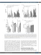
Figure 3. Natural estrogens affect human hematopoietic stem and progenitor cells differently. (A) Total number of estrogen-treated hematopoietic stem and pro- genitor cells (HSPC) after 4 days in culture. Different concentrations (10 nM, 100 nM, 1 mM, 10 mM, 100 mM and 500 mM) of the natural estrogens (E1, E2, E3 and E4) were used. (B) Total number of hematopoietic stem cells (HSC: hCD34+hCD38-hCD90+hCD45RA-) after 4 days in culture. Different concentrations (10 nM, 100 nM, 1 mM, 10 mM, 100 mM and 500 mM) of the natural estrogens (E1, E2, E3 and E4) were used. (C) Cell cycle analysis of HSPC treated with 100 nM E2 or E4. G0/G1-phase (left panel), S-phase (middle panel) and G2/M-phase (right panel). (D) Total cell number of estrogen-treated HSPC after 8 days in culture. Data were obtained from three to five biological replicates and are presented as mean ± standard deviation. Statistical significance was analyzed by one-way analysis of vari- ance with the Fisher least significant difference test: *P<0.05 and **P<0.01.
15). Treatment with E2 or E4 alone tended to increase the percentage of cells in S/G2/M-phase as previously described; however, the addition of ESR2 antagonist seemed to block the increase of cells in S/G2/M-phase induced by E4 (Online Supplementary Figure S3J). Less clear- ly, ESR1 and GPER1 antagonists seemed to reduce the number of cells in S/G2/M-phase in E2-treated HSPC. We also assessed the expression of ESR1 and ESR2 in human HSPC cultured with estrogens for 4 days by immunofluo- rescence analysis (Online Supplementary Figure S3K and L). While ESR1 fluorescence intensity increased slightly but significantly with 100 nM of E2 or E4 (Online Supplementary Figure S3K and M), ESR2 expression was significantly increased in the presence of both E2 and E4 (Online Supplementary Figure S3L and N). Moreover, estro-
gen treatment increased the percentage of human HSPC showing a polarized localization of ESR1 at the membrane (Online Supplementary Figure S3K and O). Furthermore, the treatment with estrogens enhanced the percentage of hCD34+ cells with cytoplasmic localization of ESR2 (Online Supplementary Figure S3L and P).
Collectively, these data indicate that natural estrogens regulate human HSPC through the signaling of estrogen receptors.
E2 and E4 increased the number of human hematopoietic stem and progenitor cells in an in vitro model of human hematopoietic niche
Subsequently, we investigated the indirect effect of E2 and E4 on HSPC in an in vitro model of the human
haematologica | 2021; 106(6)
1663
