
Page 159 - 2021_06-Haematologica-web
P. 159
E4 enhances human HSPC engraftment in NSG mice
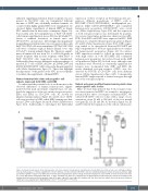
enhanced engrafting potential in female recipients was also present in CB-CD34+ cells, we transplanted different amounts of HSPC into sublethally irradiated animals. As occurred when highly purified HSC were transplanted, we observed higher engraftment of human HSPC in female NSG animals than in their male counterparts (Figure 1A). Four months after the transplantation of 5x10⁴ CB-CD34+ cells, human engraftment in mouse BM was 61.06±26.07% (mean ± standard deviation) in female mice and 18.94±13.93% in male mice. Interestingly, this impairment in engrafting potential in males was even greater when only 5x10³ CB-CD34+ cells were transplanted (38.74±30.42% BM cells were of human origin in female animals versus only 0.19±0.27% in male animals) (Figure 1B). Therefore, engraft- ment of human cells was from 3.2- to >200-fold greater in female recipients than in male recipients when 5x10⁴ or 5x10³ CB-CD34+ cells, respectively, were transplanted. Additionally, there were no differences in the percentages of myeloid, B, T cells or HSPC (hCD34+, hCD34+hCD38- and hCD34+hCD38-hCD90+ cells) between the human engrafted cells (Online Supplementary Figure S1). These data highlight the importance of the gender of the NSG mouse recipients to facilitate the engraftment of human HSPC.
Human hematopoietic stem and progenitor cell subsets expressed both ESR1 and ESR2
To understand the potential role of sex hormones in the observed differences of human hematopoietic engraft- ment between male and female recipient mice, we ana- lyzed the expression of the two main estrogen receptors, ESR1 and ESR2, in CB-CD34+ cells. As shown by immunostaining analysis (Figure 2A and B), most CD34+ cells were positive for ESR1, while ESR2 staining was dim- mer in CD34+ cells (Figure 2A and B; Online Supplementary Figure S2A). Additionally, to investigate the differential
expression of these receptors in the hematopoietic pro- genitors, different populations of HSPC, such as HSC/MPP (CD34+CD38-CD45RA-), multilymphoid pro- genitors (MLP, CD34+CD38-CD45RA+) and committed hematopoietic progenitors (CD34+CD38+), were sorted out (Online Supplementary Figure S2B) and the expression of both estrogen receptors was determined by quantita- tive reverse transcriptase polymerase chain reaction (qRT- PCR). Both ESR1 and ESR2 were expressed in HSC, MLP and in more committed hematopoietic progenitors (Figure 2C and D; Online Supplementary Figure S2C). ESR1 expres- sion tended to be upregulated between HSC/MPP and MLP compartments to decrease again in the most commit- ted hematopoietic progenitors (Figure 2C). In contrast, ESR2 expression seemed to follow an opposite pattern with high values in both HSC/MPP and committed hematopoietic progenitors but reduced levels in the MLP cell population (Figure 2D). In both cases, although some tendencies were observed, no statistically significant dif- ferences were documented. Like ESR1 and ESR2, the newly identified estrogen receptor, GPER1, was also detected by RT-PCR in CB-CD34+ cells from different donors (Online Supplementary Figure S2D). Consequently, human HSPC might respond to natural estrogens through any of the estrogen receptors.
Natural estrogens modified human hematopoietic stem and progenitor cells in vitro
Once we had demonstrated that both estrogen recep- tors were expressed in HSPC, we wanted to investigate a potential direct effect of estrogens on human HSPC. We cultured CB-CD34+ cells for 4 days with a range of con- centrations, from 10 nM to 500 mM, of the four natural estrogens (E1, E2, E3 and E4). As shown in Figure 3A, E1 and E3 reduced the expansion of the cells in culture prac-
AB
Figure 1. Human hematopoietic stem and progenitor cells show superior hematopoietic engraftment in female NSG mice than in male ones. (A) Representative flow cytometry analyses of human engraftment of 5x10⁴ umbilical cord blood CD34+ (CB-CD34+) cells into sublethally irradiated female (left panel) and male (right panel) NOD.Cg-Prkdcscid Il2rgtm1Wjl/SzJ (NSG) mice 4 months after transplantation. (B) Percentage of human hematopoietic cells, hCD45+, in the bone marrow of female (F) or male (M) animals transplanted with 5x10³ or 5x10⁴ CB-CD34+cells. Data were obtained from six independent biological replicates and are presented by dots and box-plots that represent the interquartile range (p75, upper edge; p25, lower edge; p50, midline; p95, line above the box; and p5, line below the box). Statistical significance was analyzed by the Mann-Whitney U test; **P<0.01 and ****P<0.001.
haematologica | 2021; 106(6)
1661
