
Page 162 - 2021_06-Haematologica-web
P. 162
S. Fañanas-Baquero et al.
ABC
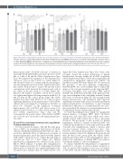
Figure 4. The impact of E2 and E4 on hematopoietic stem and progenitor cells in an in vitro model of the human hematopoietic niche. (A) Total hematopoietic cells after 1 week of co-culture with human bone marrow mesenchymal stromal cells (BM-MSC) in the presence of estrogens. (B) Total hCD34+ cells after 1 week of co-culture with human BM-MSC in the presence of estrogens. (C) Colony-forming units (CFU) derived from hematopoietic stem and progenitor cells after 1 week of co-culture with human BM-MSC in the presence of estrogens. Data were obtained from three to six biological replicates and are presented as the mean ± standard deviation. The statistical significance was analyzed by one-way analysis of variance with the Fisher least significant difference test: *P<0.05 and **P<0.01.
hematopoietic niche. CB-CD34+ cells were co-cultured on an irradiated human BM-MSC layer in the presence of 100 nM or 1 mM of E2 and E4 (Online Supplementary Figure S4A). We analyzed the expansion of the hematopoietic cells in two ways: (i) after 1 week of co-culture (Figure 4), and (ii) after 4 weeks of co-culture with the estrogen pres- ent only during the first week (Online Supplementary Figure S4CandD).From10nMto1mMofE4andthelowest concentration of E2 increased the hematopoietic cells in the culture in the first week of co-culture (Figure 4A). Likewise, the number of hCD34+ cells in the co-culture was significantly higher following treatment with E4 or 10 nM E2 than in the control group (Figure 4B; Online Supplementary Figure S4B). However, we could not detect significant differences in the functionality of the hCD34+ cells in CFU assays (Figure 4C). Furthermore, the effect of these two estrogens on the expansion of human hematopoietic cells or hCD34+ cells did not seem to be enhanced after 4 weeks in co-culture with an initial single dose (Online Supplementary Figure S4C and D). Consequently, the positive effect of E2 and E4 on HSPC also occurs in an in vitro model of the human hematopoi- etic niche.
E2 and E4 boosted human hematopoietic engraftment in immunodeficient mice
To better evaluate the impact of E2 and E4 on the prop- erties of HSPC, we transplanted 5x10⁴ human CB-CD34+ cells into sublethally irradiated male NSG mice, in order to avoid any additional effects of endogenous estrogens of female recipient mice, and 3 days later treated the animals with vehicle or daily low doses of either E2 or E4 (2 mg of estrogen per day) for 4 days (Figure 5A). Human hematopoietic engraftment was evaluated in the mouse BM by FACS analysis 4 months after transplantation (Online Supplementary Figure S5A). Surprisingly, the human hematopoietic contribution was significantly higher in the estrogen-treated animals than in vehicle-treated ones
(Figure 5B; Online Supplementary Figure S5A). None of the estrogens altered the normal distribution of human hematopoietic lineages within the hCD45+ population (Online Supplementary Figure S5B-D). More importantly, E4 administration significantly enhanced the hCD34+ cell population in male NSG mice (Figure 5C). No increase in the presence of the more primitive compartment, hCD34+hCD38-, was observed (Figure 5D). To explore the impact of the estrogen treatment on the long-term HSC, secondary transplants were performed. One million hCD45+ cells, purified from the BM of the primary recipi- ents, were transplanted into sublethally irradiated female NSG mice. As shown in Figure 5E, the estrogen-treated human hematopoietic cells maintained their long-term engraftment potential without any observable problem in human hematopoietic reconstitution or any abnormal proliferation. This led us to conclude that these two estro- gens, particularly E4, enhance in vivo human hematopoiet- ic engraftment in male immunodeficient mice.
To study this finding in more depth, we transplanted limited numbers of human HSPC (5x10³ CB-CD34+ cells/mouse), into male NSG mice, which were subse- quently treated with vehicle, E2 or E4 as previously described. The percentage of mice positive for human engraftment, defined as animals in which hCD45+ cells constituted more than 0.1% of the cells in the mouse BM 4 months after transplantation, tended to increase after estrogen treatment (Online Supplementary Figure S5E). Moreover, the human hematopoietic chimerism of the positive animals seemed to be higher in the group treated with E4 than in the group given the vehicle (Online Supplementary Figure S5F). So, E2 and E4 might be able to improve the engraftment of human HSPC even when a very limited number of cells are transplanted.
To explore whether the enhancement of engraftment mediated by estrogens occurred in female recipients as well, we repeated the transplant of this very low number of CB-CD34+ cells into sublethally irradiated female NSG
1664
haematologica | 2021; 106(6)
