
Page 125 - 2021_06-Haematologica-web
P. 125
Regulation of F8 promoter activity
Ets-1 and Ets-2 in vitro co-operation in F8 promotor regulation
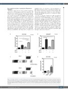
In order to explore the role of Ets-1 and Ets-2 in pF8 reg- ulation, the -1,175 bp region of pF8 was cloned into the Nanoluc vector, and pF8 activity assessed in the presence of Ets-1, Ets-2 or in combination. Two human cell lines were transfected: ECV-304 and HEK293T. The ECV-304, previously considered as spontaneously transformed human umbilical endothelial cells,32 were selected as a model expressing FVIII, Ets-1 and Ets-2 at the mRNA and protein level, while HEK293T as cells expressing low lev- els of Ets-1 and Ets-2 (Figure 1C and D). The luciferase assays showed different but consistent results between the two cell types tested. A 3- and 5-fold upregulation was observed in ECV-304 upon co-transfection of pF8 with Ets-1 and Ets-1/Ets-2, respectively (Figure 2A). No signifi- cant effect was observed with Ets-2 alone. This data high- lights that Ets-1 appears to play a major role on pF8 trans- activation. In HEK293T cells where Ets-1 is expressed at
negligible levels, we observed a 7-fold up-regulation of pF8 only in response to Ets-1 overexpression, with no fur- ther increase observed in presence of Ets-2 (Figure 2B). These results reinforce the central role of Ets-1 in regulat- ing pF8 and highlight the presence of cell-specific regula- tory mechanisms.
In order to elucidate whether the pF8 up-regulation observed in ECV-304 in response to Ets-1 and Ets-2 is mediated by a direct DNA binding of Ets, we generated Ets mutants carrying an in-frame deletion of their DNA binding domain (DBD) (Figure 2C). We evaluated their co- operation with the wild-type (WT) counterparts in the regulation of pF8 activity. Ets-1 without DBD failed to upregulate the pF8, while Ets-1 co-expressed with the Ets2-DBD mutant, preserved the 5-fold increase in pF8 activity, as observed with the WT proteins (Figure 2D). These results suggest that in the regulation of pF8, Ets-1 may directly bind the DNA, while Ets-2 modulates pF8 through its interaction with Ets-1.33
AB
CD
Figure 2. In vitro Ets-1 and Ets-2 co-operation in F8 promotor transactivation. (A and B) Histograms report the fold increase of luciferase activity after transfection of F8 promotor (pF8) alone (set as 1) or in combination with Ets-1, Ets-2 or both in (A) ECV-304 or (B) HEK293T cell lines. (C) Schematic representation of Ets-1 and Ets-2 protein structure, with highlighted amino acid position of the DNA binding domain (DBD). For both proteins, TAD indicates the transactivation domain while ID indicates the inhibitory domain. (D) Histograms report the fold increase of luciferase activity after transfection of pF8-1175 alone (set as 1) or with the combination of Ets-DBD with the non-mutated counterparts. Results are expressed as mean ± standard deviation from three independent experiments performed in triplicate. *P<0.05.
haematologica | 2021; 106(6)
1627
