
Page 284 - 2021_05-Haematologica-web
P. 284
CASE REPORTS
Acute leucoencephalomyelopathy and quadripare-
sis after CAR T-cell therapy
Chimeric antigen receptor (CAR) T-cell therapy maybe associated with neurologic toxicity, also referred to as immune effector cell-associated neurotoxicity syndrome (ICANS),1,2 that typically manifests as encephalopathy. Here, we report two patients, with no known prior neuro- logical disease, treated on ZUMA-1 trial, who developed acute leucoencephalomyelopathy with quadriparesis after treatment with axicabtagene ciloleucel (axi-cel).3,4
Patient 1 is a 41-year-old female with refractory diffuse large B-cell lymphoma after four lines of systemic therapy including high-dose chemotherapy plus autologous stem cell transplantation. She had bulky nodal and splenic dis- ease (Online Supplementary Figure S1A) prior to treatment with axi-cel.3,4 Her baseline C-reactive protein (CRP) and ferritin were elevated at 136 mg/L and 9,821 ng/mL, respectively (Figures 1A and B). She experienced intermit- tent grade 1 cytokine release syndrome (CRS) with fever and tachycardia from days 1-6.5 On day 2, she developed grade 3 ICANS with confusion concurrently with fever, which resolved promptly with tocilizumab and dexam- ethasone administration along with a dose increase of lev- etiracetam that was started on day 0 for seizure prophylax- is. On day 4, grade 3 ICANS recurred with aphasia which was non-responsive to a second dose of tocilizumab. On
day 5, she developed grade 4 ICANS with clonic seizures evolving to status epilepticus requiring ventilator support, additional anti-epileptic medications, and high-dose methylprednisolone. On days 6 and 7, she had two gener- alized tonic-convulsive seizures with further electrograph- ic seizures (Online Supplementary Figures S5-6). The seizures were eventually controlled with lorazepam, phenytoin, levetiracetam, and phenobarbital. On day 8, as patient mental status improved, she was noted to be weak in the lower extremities with rapid progression to quadri- paresis, mute plantar reflexes, and lack of bladder control. Cerebrospinal fluid (CSF) analysis showed an increased protein level but no evidence of infection. Magnetic reso- nance imaging (MRI) of the brain and spine on day 9 showed findings concerning for acute leucoen- cephalomyelopathy with symmetrical T2 hyperintensity within the centrum semiovale with sparing of the U-fibers, the superior cerebellar peduncle and striking dif- fuse cerebral edema (Figure 2A). There was involvement of the diffuse periventricular white matter, external capsule, and posterior limb of the internal capsule, and posterior limb of the internal capsule. MRI of the spine demonstrat- ed centromedullary holocord involvement (Figure 2A). By day 11, upper extremity strength started to improve and she self-extubated. Her cognitive function quickly improved but she experienced retrograde amnesia span- ning a time period of about 2 weeks prior to this event. By
AD
BE
CF
1504
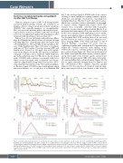
Figure 1. Clinical and biological parameters, and therapeutic intervention over time in patient 1 (A, B, C) and 2 (D, E, F). Conditioning chemotherapy with flu- darabine and cyclophosphamide was given on days -5 to -3, with infusion of axicabtagene ciloleucel Chimeric antigen receptor (CAR) T cells on day 0. Tocilizumab was administered intravenously at 8 mg/kg, dexamethasone intravenously at 10 mg every 6 hours, and methylprednisolone intravenously at 1,000 mg/day with gradual taper. The CAR T-cell levels in the peripheral blood in patients 1 (C) and 2 (F) were compared to the median CAR T-cell levels in patients treated on the corresponding cohort of ZUMA-1 study (cohorts 1 and 2 for patient 1 and cohort 4 for patient 2). HR: heart rate; Tmax: temperature maximum, CRP: C-reactive protein.
haematologica | 2021; 106(5)
