
Page 282 - 2021_05-Haematologica-web
P. 282
Letters to the Editor
CD80, CD86 and CD154, as these antigens were barely expressed on leukemic B cells (data not shown).
With regard to the main biological prognostic factors in CLL, no significant differences in antigen modulation were observed according to the IGHV status. This finding is in agreement with the evidence that CLL patients with unmutated or mutated IGHV genes respond equally well to treatment with ibrutinib1 and supports the hypothesis that peripheral lymphocytosis is more related to the role of BTK in cell adhesion and migration, and less to its function in BCR signaling.11 On the contrary, the presence of TP53 mutations was associated with a lower down- modulation of CD62L MFI (21/119 TP53 mutated vs. 98/119 TP53 wild-type cases, DMFI: 202±172 vs. 479±424, P=0.039). Taking into account a lesser depend- ency of TP53-mutated CLL cells on the BCR pathway for survival and proliferation,12 these data may help to eluci- date the biological basis for the suboptimal response of TP53-disrupted patients to ibrutinib alone or in combina- tion with anti-CD20 monoclonal antibodies.13,14
A differential response was also observed with regard to cytogenetic aberrations. On D14, CLL samples harbor- ing an isolated trisomy 12 (tris12+ CLL, n=21/119) showed a significantly greater downmodulation of CD49d and CD62L when compared to CLL cases with other fluorescence in situ hybridization patterns, with the exception of CD49d in del17p+ cases (Figure 2A). These results reflected the increased expression of integrins on tris12+ CLL cells. A higher baseline MFI was indeed observed for CD49d (tris12+ vs. normal karyotype, P=0.016; tris12+ vs. del13q14+, P=0.002; tris12+ vs. del11q+, P=0.0003; tris12+ vs. del17p+, P=0.029) and CD62L (tris12+ vs. del11q+, P=0.003; tris12+ vs. del17p+, P=0.026) compared to tris12– samples, which may clarify the specific tropism of tris12+ cells towards lymph nodes
and the shortened lymphocytosis observed during ibruti- nib monotherapy.15
Lymphocytosis was observed in 36 of the 119 CLL patients (30.3%), with D0 vs. D14 absolute lymphocyte counts being 42.6±26.4x10⁹/L vs. 92.8±57.3x10⁹/L, respectively (P<0.00001). The lymphocytosis appeared associated with the downmodulation of at least four anti- gens (P=0.050), independently of the type of antigens involved. No significant differences in antigen modula- tion were recorded according to the presence (36/119 cases) or absence (83/119 cases) of lymphocytosis, sug- gesting that ibrutinib-mediated inhibition of microenvi- ronmental interactions could not always turn into an increase in absolute lymphocyte count, probably because of the cytotoxic effect on both tissue-resident and mobi- lized CLL cells. Accordingly, when CLL patients were subdivided on the basis of level of CD20 expression into those with higher (CD20high MFI ≥1000, n=62/119) or lower (CD20low MFI <1000, n=57/119) expression, CD20high cases showed both a greater clearance of CLL cells (CLL median absolute cell counts on D14 in CD20high vs. CD20low cases: 38.9±46.7x10⁹/L vs. 58.3±48.6x10⁹/L, P=0.030) (Figure 2B) and a lower incidence of lymphocy- tosis (15/62 [24.6%] vs. 21/57 [36.8%], P=0.083) on D14. In addition, CD20high CLL samples were characterized by a significantly greater downmodulation of CD38, CD44, CD49d and CD62L antigens (Figure 2C), suggesting that these cells may be more prone to mobilize into the periphery and be exposed to monoclonal antibody-medi- ated killing.
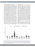
Finally, in order to assess a possible relationship between antigen expression changes and response to treatment, the MFI modulation on D14 was correlated to the amount of residual CLL cells detected in the periph- eral blood 8 months after the start of IR treatment.
Figure 3. Correlation between the changes of expression of adhesion molecules, chemokine receptors and activation markers on day 14 compared to base- line and chronic lymphocytic leukemia absolute cell counts observed at 8 months after the start of ibrutinib plus rituximab treatment. Chronic lymphocytic leukemia absolute cell counts were calculated from flow cytometric minimal residual disease values available for 91 of the 119 patients included in the study. Data are presented as mean fluorescent intensity (MFI) of surface marker expression obtained after 14 days (D14) of ibrutinib plus rituximab treatment with respect to baseline (D0) (ΔMFI: D14MFI-D0MFI; **P<0.01, *P<0.05, unpaired Student t-test). The antigen expression changes observed in the totality of sam- ples analyzed for each antigen are added for comparison.
1502
haematologica | 2021; 106(5)
