
Page 285 - 2021_05-Haematologica-web
P. 285
Case Reports
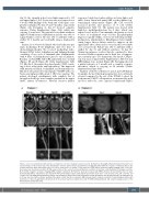
day 16, the strength in her lower limbs improved to 3/5 and upper limbs to 5/5. Corticosteroids were tapered over 3 weeks. MRI findings of the brain and of the spine com- pletely normalized by day 27 and 6 months, respectively (Figure 2A). Restaging on day 28 showed complete response (Online Supplementary Figure S1A), which was ongoing 3 years later. The patient’s lower limb weakness improved with intense rehabilitation and she was able to regain bladder control. She was able to ambulate with a walker by 6 months and eventually using a single-point cane.
Patient 2 is a 30-year-old female who had refractory pri- mary mediastinal B-cell lymphoma after two lines of chemoimmunotherapy. She received mediastinal radio- therapy (20Gy) before leukapheresis and bridging therapy consisting of one cycle of rituximab plus bendamustine with 4 days of dexamethasone prior to axi-cel infusion. Baseline cerebral MRI, CSF, CRP, and ferritin were normal (Figures 1D and E; Figure 2B; Online Supplementary Table S1). On day 2, the patient developed grade 2 CRS consist- ing of fever, tachycardia, and hypotension.5 She improved with intravenous fluids, tocilizumab, and dexamethasone. On day 5, the patient developed grade 3 ICANS with con- fusion and aphasia while grade 1 CRS was ongoing. The patient developed quadriparesis with complete loss of strength in both legs, near complete paralysis in the upper limbs, urinary incontinence, bilateral extensor plantar
responses, brisk deep tendon reflexes in lower limbs, and ankle clonus. Brain and spinal MRI revealed diffuse lep- tomeningeal enhancement (Figure 2B). CSF revealed increased protein and presence of T cells (Online Supplementary Table S1). The patient was treated with high-dose methylprednisolone for 4 days, which was tapered over 2 weeks. Concomitantly, the patient received 4 doses of tocilizumab every 6 hours. Encephalopathy improved quickly within a few hours following methyl- prednisolone administration. Quadriparesis lasted signifi- cantly longer. Motor deficits started to improve in the upper limbs after 2 days and recovered in the lower limbs over several weeks. Patient was able to ambulate with a walker by day 21 and without assistance by day 28. Urinary incontinence resolved but she continued to have decreased bladder sensation and a distal loss of tempera- ture sensation up to T10 dermatome. By day 28, CSF pro- tein was near normal (Online Supplementary Table S1) and MRI findings were normal (Figure 2B). Restaging showed a partial response at 1 month and a complete response thereafter, which is ongoing at 18 months (Online Supplementary Figure S1B).
Analysis of the blood (Patient 1 and 2) and CSF (Patient 2) samples showed that many parameters were extremely elevated compared to the rest of the ZUMA-1 cohort. In both patients, CAR T-cell expansion, as measured by peak and area under the curve, appeared significantly higher
AB
Figure 2. Leucoencephalomyelopathy changes on magnetic resonance imaging in patient 1 and 2. (A) Patient 1: T2-weighted fluid attenuation inversion recov- ery axial images of the brain (top 2 rows) demonstrate evolution and resolution of symmetrical T2 hyperintensity within the superior cerebellar peduncle, the centrum semiovale white matter with respect of U-fibers, from day 9 to 6 months. T2 fast spin echo (FSE) with fat saturation sagittal imaging of the cervico-tho- racic spine (row 3) and representative T2 FSE axial sections of the cervical cord (row 4) illustrate resolution of extensive centromedullary T2 changes over time. By 6 months, magnetic resonance imaging (MRI) changes normalized. (B) Patient 2: brain and cervical spine MRI performed at neurotoxicity onset on day 5 demonstrating diffuse infratentorial and supratentorial leptomeningeal uptake predominant around the brain stem, cervical spinal cord, and cerebellar and occipital sulci (3D FLAIR with gadolinium). Diffuse leptomeningeal enhancement (3D T1 Black Blood with gadolinium) and T2 hyperintensity extending from C2 to C4 was also noted with a slender swelling of the spinal cord. At day 28 re-evaluation, MRI showed almost complete resolution of leptomeningeal uptake with persistence of discreet gadolinium enhancement in the posterior parieto-occipital sulcus (3D T1 VISTA with gadolinium) and complete disappearance of the intramedullary T2 hyperintensity.
haematologica | 2021; 106(5)
1505
