Page 187 - 2021_05-Haematologica-web
P. 187
TAK1 inhibition in myeloma
AB
C
D
E
FG
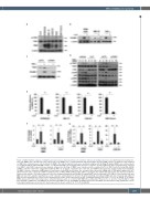
Figure 3. TAK1 inhibition suppresses VCAM-1 expression in bone marrow stromal cells and their adhesion to multiple myeloma cells. (A) Human bone marrow stro- mal cells (BMSC) were expanded in 6-well culture plates. The BMSC were cocultured with the indicated multiple myeloma (MM) cell lines for 24 hours. After washing out MM cells, cell lysates were collected from the BMSC. The indicated protein levels were examined using western blotting. (B) Human BMSC were cultured alone or cocultured with the indicated MM cell lines, or cultured with TNF-α at 10 ng/mL in the presence or absence of LLZ (5 mM) for 2 days. Cell lysates were collected from BMSC, and VCAM-1 expression was analyzed using western blotting. (C) BMSC were transduced with scrambled (siCTL) or TAK1 small interfering RNA (siRNA) (siTAK1), and then cultured for 2 days with or without TNF-α at 10 ng/mL. Cell lysates were collected, and VCAM-1 expression was analyzed using western blotting. (D) BMSC cells were starved in α-MEM with 1% fetal bovine serum (FBS) for 12 hours. The cells were then cultured in α-MEM with 1% FBS with or without LLZ at 5 mM for 3 hours (left), or transduced with scrambled (siCTL) or TAK1 siRNA (siTAK1) (right). TNF-α at 10 ng/mL was added and cells lysates were harvested after the indicated time periods. The indicated protein levels were analyzed using western blotting. (E) Human BMSC were treated with LLZ (5 mM) for 1 day, and the indicated MM cells were then added in quadruplicate at 105 cells/well, and incubated for 4 hours. By gentle pipetting, non-adherent MM cells were removed, and adherent MM cells were quantitated in a fluorescence multi-well plate reader. Data represent the means ± standard deviation (SD) (n=4). *P<0.05, by ANOVA. (F, G) Human BMSC prepared in 6-well culture plates were cultured in triplicate alone or cocultured with MM cells as indicated in the presence or absence of LLZ (5 mM) for 1 day. After washing out MM cells, total RNA was isolated from the BMSC. IL-6 (f) and RANKL (g) mRNA expression in the BMSC was determined using quantitative reverse transcription polymerase chain reaction. Data represent the means ± SD (n=3). *P<0.05, by ANOVA.
haematologica | 2021; 106(5)
1407


