
Page 189 - 2021_05-Haematologica-web
P. 189
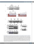
TAK1 inhibition in myeloma
A
B
C
DE
F
Figure 5. TAK1 inhibition restores osteoblastogenesis suppressed by multiple myeloma cells as well as major inhibitors for osteoblastogenesis in multiple myelo- ma. (A) Conditioned media from the indicated multiple myeloma (MM) cell lines (MM CM) at 25%, or cytokines including TNF-α (1 ng/ml), TGF-b (10 ng/ml), IL-3 (10 ng/ml), IL-7 (10 ng/mL), or activinA (10 ng/mL) were added onto cultures with the MC3T3-E1 cells. After culturing for 24 hours, cell lysates were collected, and phos- phorylated TAK1 (p-TAK1) and TAK1 levels were analyzed using western blotting. β-actin served as a loading control. (B) MC3T3-E1 cells were cultured in the presence or absence of LLZ (0.3 mM) in osteogenic media with BMP-2 (25 ng/mL) in 24-well culture plates. MM cells CM from the indicated cell lines and primary MM patient were added at 25%. TNF-α (1 ng/mL), TGF-b (10 ng/mL), IL-3 (10 ng/mL), IL-7 (10 ng/mL), or activinA (10 ng/mL) were added to the indicated wells. After culturing for 14 days, the cells were fixed and mineralized nodule formation was visualized using Alizarin red staining. (C) MC3T3-E1 cells were cultured for 4 days with MM CM (25%) or TNF-α (1 ng/mL) in the presence or absence of LLZ (0.3 mM) in osteogenic media with BMP-2 (25 ng/mL). Then, cell lysates were collected and Osterix (OSX) expression was assayed using western blotting. (D) MC3T3-E1 cells were starved in α-MEM with 1% FBS for 12 hours, and then treated with or without LLZ at 0.3 μM for 3 hours, or transfected with scrambled (siCTL) or TAK1 (siTAK1) small interfering RNA. TGF-b was added at 10 ng/mL, and cell lysates were harvested after the indicated time periods. The expression of indicated proteins were analyzed using western blotting. (E) (Upper) MC3T3-E1 cells were treated with LLZ at the indicated concentrations for 12 hours. (Lower) MC3T3-E1 cells were treated with LLZ for 30 minutes prior to adding TGF-b (10 ng/mL), then cultured for 12 hours. The expression of Smad6 protein was analyzed using western blotting. (F) MC3T3-E1 cells were starved with or without LLZ at 0.3 mM for 12 hours, or transfected with scrambled (siCTL) or TAK1 (siTAK1) siRNA. BMP-2 was added at 25 ng/mL, and cell lysates were harvested after the indicated time periods. The expression of indicated proteins were analyzed using western blotting.
haematologica | 2021; 106(5)
1409
