
Page 185 - 2021_05-Haematologica-web
P. 185
TAK1 inhibition in myeloma
A
BCD
E
FG
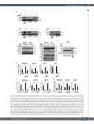
Figure 2. TAK1 inhibition abolishes IL-6- and TNF-α-induced signaling in multiple myeloma cells. (A) Human bone marrow stromal cells (BMSC) were expanded in 24-well culture plates. The indicated multiple myeloma (MM) cell lines were cultured alone or cocultured with BMSC (upper), or cultured in the presence or absence of IL-6 (lower left) or TNF-α (lower right) for 24 hours (h), and cell lysates were then collected. PIM2 expression was analyzed using western blotting. b-actin was blot- ted as a loading control. (B, C) RPMI8226 cells were cultured in α-MEM with 1% FBS for 12 h for serum starvation. The starved cells were then cultured in α-MEM with 1% fetal bovine serum (FBS) with or without LLZ1640-2 (LLZ) at 3 mM. Three hours later, IL-6 (b) or TNF-α (c) at 10 ng/mL was added. After the indicated time periods, cell lysates were collected. The expression of phosphorylated TAK1 (p-TAK1), TAK1, phosphorylated STAT3 (p-STAT3), STAT3, phosphorylated IκBα (p-IκBα), IκBα, phosphorylated p38MAPK (p-p38), p38MAPK (p38), phosphorylated ERK (p-ERK), and ERK were detected using western blotting. b-actin was used as a loading control. (D) RPMI8226 cells after starvation were cultured in α-MEM with 1% FBS with or without LLZ (5 mM) or the PIM inhibitor SMI-16a (50 mM). Three hours later, TNF-α at 10 ng/mL was added as indicated. After incubating for 15 minutes, whole lysates and nuclear fractions were extracted. The protein levels of p65 were ana- lyzed using western blotting. Glyceraldehyde 3-phosphate dehydrogenase (GADPH) and p84 were used as protein loading controls for cytoplasmic and nuclear pro- teins, respectively. (E) The indicated MM cell lines were cultured alone or cocultured with human BMSC expanded in 24-well culture plates for 24 hours in the pres- ence or absence of LLZ at 5 mM. MM cells were then collected, and their viability was analyzed using a WST8 assay. (F) The indicated MM cell lines were transduced with scrambled (siCTL) or TAK1 siRNA (siTAK1), cultured alone or cocultured with human BMSC expanded in 24-well culture plates for 24 hours. MM cells were then collected, and their viability was analyzed using a WST8 assay. (G) Indicated MM cells were cocultured for 24 hours with or without human BMSC which were trans- duced with scrambled (siCTL BMSC) or TAK1 siRNA (siTAK1 BMSC). MM cells were then collected, and their viability was analyzed using a WST8 assay.
haematologica | 2021; 106(5)
1405
