
Page 86 - 2021_04-Haematologica-web
P. 86
T. D’Altri et al.
AC
B
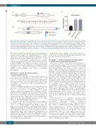
Figure 1. Generation of the Asxl1G643W mutant mouse line. (A) Schematic representation of the three last exons of the Asxl1 gene. The red star represents the G643W mutation. The “g” inserted in the mutated allele is indicated in red. The protein sequence is in capitals and the asterisk represents the stop codon generated as a result of the frameshift. The blue arrows represent primers used for genotyping (two forward primers have been used to selectively anneal the wild-type [WT] and mutated sequence, respectively). (B) Schematic representation of WT and G643W mutant ASX1 proteins. The major plant homeodomain (PHD) and additional sex combs homology domain (ASXH) are indicated. Amino acids are numbered. The light blue arrows represent the primers used for quantitative RT-PCR. (C) The relative expression of Asxl1 cDNA in Asxl1+/+, Asxl1G643W/+ and Asxl1G643W/G643W BM cells, determined by quantitative RT-PCR (n=3 mice in each experimental group).
12 codons downstream, thereby precisely mimicking the human situation (Figure 1A). The mutated Asxl1G643W allele expresses a truncated form of ASXL1 lacking the C-termi- nal plant homeodomain (Figure 1B). Both Asxl1G643W/+ and Asxl1G643W/G643W mice express elevated levels of Asxl1, demonstrating that the mutated allele escapes nonsense- mediated mRNA decay (Figure 1C). This is in line with the previously observed expression of truncated ASXL1 in patient cells.6 The increased levels of the mutated mRNA could potentially be the result of a feedback mechanism.
The Asxl1G643W variant has minimal impact on normal hematopoiesis
We next assessed the impact of the G643W mutation in the context of normal hematopoiesis. Both Asxl1G643W/+ and Asxl1G643W/G643W mice were born at the expected Mendelian ratios and showed a normal lifespan. This suggests that the ASXL1G643W variant has no impact on embryonic devel- opment or aging.
We next analyzed peripheral blood for the relative fre- quencies of the major blood lineages. Six-month old Asxl1G643W/+ and Asxl1G643W/G643W mice displayed no major changes within the peripheral blood compared to wild- type (WT) controls (Figure 2A). However, at 18 months, we observed a skewing towards the myeloid lineage in ASXL1 mutated mice (Figure 2B-C, Online Supplementary Figure S1A). The age-dependent skewing was accompa- nied by a mild splenomegaly (Figure 2G).
Consistent with the lack of changes in the peripheral blood in young mice, 6-month old Asxl1G643W/+ and Asxl1G643W/G643W neither displayed any changes within the hematopoietic stem cell (HSC) and multipotent progenitor (MPP) compartment, nor within the distribution of mature blood lineages in the bone marrow (Figure 2D-F, Online Supplementary Figure S1B). Competitive transplantation of ASXL1-mutated BM cells revealed a significant but minor reduction in the ability of Asxl1G643W/G643W BM to reconsti- tute hematopoiesis, suggesting that HSC functionality is only mildly affected (Figure 2H).
Collectively, these findings show that mutation of ASXL1 leads to a mild and age-dependent perturbation of the hematopoietic system in mice.
The ASXL1G643W variant accelerates the development of CEBPA mutant acute myeloid leukemia
Mutations in ASXL1 and CEBPA are frequently co- occurring in AML patients.23,24 In order to test the potential functional interplay between these factors, we crossed all three Asxl1 genotypes onto either a Cebpafl/p30; Mx1Cre or Cebpafl/+; Mx1Cre background resulting in a total of six genotypes. We subsequently transplanted BM from these mice into lethally irradiated recipients and 3 weeks later induced the deletion of the conditional Cebpa allele by injections with polyinosinic:polycytidylic (pIpC) acid (Figure 3A). This strategy facilitates the deletion of the full-length Cebpa allele, thereby allowing the Cebpap30 allele to exert its oncogenic function. Consistent with pre- vious findings, Asxl1+/+; CebpaD/p30 donor cells sustain the development of AML with a median latency of 43 weeks (Figure 3B).26 Interestingly, both heterozygous and homozygous expression of the ASXL1G643W variant signifi- cantly accelerated CEBPA mutant driven AML develop- ment, with median disease latencies of 37 and 38 weeks, respectively. In contrast, none of the control CebpaD/+ geno- types lead to AML, irrespective of their Asxl1 mutation status.
Given that Asxl1G643W/G643W animals, compared to their heterozygous counterparts, displayed a somewhat more pronounced phenotype during steady-state hematopoiesis, we decided to focus on this genotype in the context of CEBPA mutant AML. Whereas Asxl1+/+; CebpaD/p30 and Asxl1G643W/G643W; CebpaD/p30 leukemias appeared morphologically identical, the latter displayed a trend towards increased levels of c-Kit (Figure 3C-E, Online Supplementary Figure S2). This suggests that the Asxl1 mutation, at least in the context of CEBPA mutant AML, could result in a slightly more immature leukemic pheno- type which would be consistent with the more aggressive nature of ASXL1 mutant AML.
1002
haematologica | 2021; 106(4)
