
Page 291 - 2021_04-Haematologica-web
P. 291
Letters to the Editor
β-thalassemia patients
gesting that either the global gene expression profile was very similar between the two types of the disease or that substantial variability in expression did not allow the identification of consistent changes, or both (Online Supplementary Table S6). In general, only small changes were detected when TM and TI were directly compared (Figure 1B) and the increased gene expression variability seen in TI patients, which did not allow the identification of the same number of significantly DEG as in the case of TM patients, also hindered the identification of DEG between TI and TM patients. Increased gene expression variability in TI potentially reflects the high level of phe- notypic heterogeneity for TI patients.
Nonetheless, the expression profiles accurately por- trayed the clinical observations of β-thalassemia. The severe TM phenotype was associated with induction of organismal injuries, as well as inhibition of key hemato- logical system genes and inflammatory response mole- cules compared to the less severe type of the disease (TI) (Figure 1C-D; Online Supplementary Table S7). Moderate changes were seen in the expression levels of various glo- bin and other interacting proteins in TI patients, whereas in TM patients the data portrayed the marked repression of β-thalassemia-related proteins with concomitant up- regulation of other globin proteins as a means of com- pensating for the ineffective erythropoiesis (Online Supplementary Figure S1). Focusing on molecular path- ways affected by the disease, gene set enrichment analy- sis (GSEA) revealed very similar pathways in both TI and TM patients as differentially represented when compared to healthy participants, in accordance with the gene expression profiles (Online Supplementary Figure S2). Several of the pathways identified have been previously linked to β-thalassemia validating our results, such as the impaired packaging of telomere ends,4 impaired unfolded protein response (UPR) pathway5 and lipid abnormalities.6,7 Nonetheless, the lack of significant changes between TI and TM patients in terms of global gene expression profiles or molecular pathways suggest that a continuous spectrum describes the disease and not distinct conditions.
We then searched for other biological confounders that could affect global expression patterns allowing patient stratification, since the study was designed to limit as much as possible all technical sources of variation (bal- anced groups in terms of sex and age, standardized cell culture protocol in all centers with the matched samples being cultured at the same time, library construction per-
Sex-specific transcriptional profiles identified in
β-thalassemia comprises a group of heterogeneous autosomal recessive hereditary anemias characterized by the reduction or absence of β-globin chain synthesis, and it is a highly prevalent disease affecting 1.5% of the glob- al population.1 Three different clinical conditions are rec- ognized in patients with β-thalassemia minor (trait) being the asymptomatic form, β-thalassemia major (TM) being the most severe form of the disease and β-thalassemia intermedia (TI) presenting with variable severity. Despite extensive characterization of the genetic basis of disease pathogenesis,2 currently the classification of patients relies on the severity of symptoms and hemoglobin (Hb) F levels regardless of the underlying genotype. Thus, the aim of the study was to develop an approach for patient stratification based on gene expression, to pinpoint the targets that dictate each phenotype and to provide a framework for the development of therapeutic strategies focused on these targets. To this end, we have analyzed the gene expression profiles of TI, TM and healthy indi- viduals using RNA sequencing (RNAseq) (National Center for Biotechnology Information [NCBI], GSE117221) and we have studied the differentially expressed genes (DEG) and pathways irrespective to patient genotype. Interestingly, after analysis of various confounding factors, we identified sex differences in the patients’ expression profiles suggesting that males and females are differentially affected by β-thalassemia. Thus, taking sex into account might benefit prognosis, diagno- sis, stratification and therapeutic management of the dis- ease.
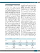
In particular, 49 subjects (after exclusion of low quality samples) were included in the analysis and organized in groups of three age- and sex-matched samples within each group (Online Supplementary Tables S1-2). RNAseq libraries were generated from erythroid precursor cell cul- tures after the isolation of peripheral blood mononuclear cells from all participants, as previously described.3 We identified 716 genes with aberrant expression between TI patients and healthy subjects, and 2,885 between TM patients and healthy subjects with most of DEG seen in TI patients being also present in TM patients when com- pared to healthy subjects, albeit with more pronounced changes (Figure 1A; Table 1; Online Supplementary Tables S3-5). However, no significantly DEG were found when TM patients were compared directly to TI patients sug-
Table 1. Numbers of significantly differentially expressed genes. Analysis
All Males Females
TI vs. H
TM vs. H
TM vs. TI
Up-regulated 147 Down-regulated 569 Total 716
315 5 1,244 9 1,559 14
Samples 16TIvs.17H 7TIvs.8H 9TIvs.9H
Samples 16TMvs.17H 8TMvs.8H 8TMvs.9H
Up-regulated Down-regulated Total
939 1,946 2,885
40 100 401 210 441 310
Samples 16TIvs.16TM 8TMvs.7TI 8TMvs.9TI Up-regulated 0 0 0 Down-regulated 0 0 1 Total 0 0 1
TI: β-thalassemia intermedia; TM: β-thalassemia major; H: healthy. Numbers of significantly differentially expressed genes are shown for all comparisons performed. The analysis is produced by DESeq2 and differentially expressed genes were defined as significant when Padj<0.1.
haematologica | 2021; 106(4)
1207
