
Page 227 - 2021_04-Haematologica-web
P. 227
Sec22b controls VWF trafficking and WPB size
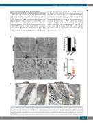
Sec22b silencing results in accumulation of von Willebrand factor in dilated rough endoplasmic reticulum Since VWF was retained in the ER, potentially along with other proteins, we used electron microscopy to examine the impact of reduced anterograde trafficking on the ER (Figure 4A). When Sec22b was silenced the rough ER (rER) appeared enlarged and ribosome-studded, mem- brane-limited, rounded structures developed, with elec- tron-dense content. These rER structures represent severe- ly dilated ER cisternae as they often retained a membra- nous connection to the rER. The dilated rER phenotype was observed in the majority of Sec22b-depleted cells (72.9%) (Figure 4B). Closer examination of the rER mor-
phology revealed that apart from the round rER structures, the luminal width of ER sheets was significantly increased in Sec22b KD cells (0.29 mm ± 0.18 mm) when compared to control cells (0.10 mm ± 0.01 mm) (Figure 4C). This sug- gests that upon removal of Sec22b the rER expands in size dramatically, possibly to facilitate the accumulation of secretory proteins such as VWF. Indeed, immunogold staining for VWF in Sec22b KD endothelial cells localized within dilated rER and was prominently found in the round, dense rER structures (Figure 4D). Taken together this shows that VWF exits the ER in a Sec22b-dependent manner and upon Sec22b silencing is retained in rER- derived structures.
AB
D
Figure 4. von Willebrand factor accumulation in dilated rough endoplasmic reticulum in Sec22b-depleted endothelial cells. (A) Electron microscopy of control and Sec22b knockdown (KD) endothelium cells (EC) (dilated rough endoplasmic reticulum [ER] shown by white arrowheads, ribosome-studded dilated ER by white aster- isks, and the connection of ER structures to rough ER sheets by a yellow arrowhead; scale bar corresponds to 2 mm). (B) Quantification of healthy versus dilated ER in control and Sec22b KD cells. (C) Quantification of ER width in control and Sec22b KD cells (t-test with Welch correction, ****P<0.0001). (D) von Willebrand factor immunogold staining (10 nm gold particles) in control and Sec22b KD EC (boxed regions are magnified on the right side with the corresponding color; scale bar rep- resents 1 mm).
C
haematologica | 2021; 106(4)
1143
