
Page 205 - 2021_04-Haematologica-web
P. 205
Oncogenic somatic mutations in plasmablastic lymphoma.
patterns of PBL.6 However EBV positivity in PBL has been found to be associated with increased expression of the programmed death ligand 1 (PD-L1) protein as well as other immune escape markers,7,8 and decreased expression of major histocompatibility class II (MHCII)/human leukocyte A (HLA)-DR molecules by the neoplastic cells.9 This has recently been found to be associated with increased antiviral cytotoxic immunity involving different immune cell populations.7
The genetic landscape of somatic mutations in PBL is unclear. So far, MYC-IGH translocations have been the most commonly detected alterations, being present in 60% of cases.10,11 Concurrent mutations in PRDM1/Blimp1 have been found in half of these cases.12 Very recently, exome sequencing of a series of HIV-positive cases of PBL showed somatic mutations involving components of the non-canonical NFκB pathway as well as genes involved in immune response,13 but the data remain limited.
Our aim was to characterize the genetic profile of a series of PBL cases using targeted exonic next-generation sequencing (NGS) and correlate the findings with EBV infection and the expression status of immune checkpoint proteins in both the population of neoplastic cells and cells in the microenvironment. In addition, we quantified the components of the microenvironment and searched for skewed T-cell populations in this tumor. We found that the mutational profile of PBL was related to EBV infection in the tumor cells and identified recurrent genetic events in MYC, STAT3 and PRDM1/Blimp1 that were more fre- quent in EBV-positive disease. In addition, we identified PD-L1 expression on tumor cells in a subset of cases as well as enrichment of tumor-associated macrophages (TAM) and programmed death 1 (PD1) reactive T cells in the microenvironment of PBL cases.
Methods
Case selection
Twenty-eight new cases were retrieved from the files of the Pathology Department of Universitario Marqués de Valdecilla Hospital (Santander, Spain), ten samples from the files of the University of Texas MD Anderson Cancer Center Hematopathology Department (Houston, TX, USA) and four cases from the Pathology Department of San Bortolo Hospital (Vicenza, Italy). Material transfer agreements were signed by the Instituto de Investigación Marqués de Valdecilla (IDIVAL) and corresponding institu- tions to share the material in the project. The study and sample collection were approved by the local ethics com- mittee (CEIC Cantabria, Institutional Review Board code 2016.168) and complied with the Declaration of Helsinki. All cases were diagnosed according to the World Health Organization (WHO) classification of Hematolymphoid Neoplasms.14 All cases had to be negative for pan-B-cell markers (CD20), HHV-8 and ALK in order to be included in the study. The phenotype of the cases was consistent with a plasma cell differentiation program.4,15 The clinical features of the cases were recorded and a summary is available in Online Supplementary Table S1.
Immunohistochemistry and in situ hybridization Immunohistochemical reactions were performed fol- lowing conventional automated procedures.Chromogenic in situ hybridization for EBV and its encoding RNA (EBER)
and fluorescence in situ hybridization (FISH) for the detec- tion of MYC rearrangements were also done.
Quantification of the cellular composition of the tumor and transcription factor abundance
The different lymphoid and histiocytic/dendritic sub- populations, identified with CD3, CD8, PD1, CD163, PD- L1 and MHCII/HLA DP/DR and the absolute number of nuclei showing expression of MYC and phospho-STAT3 (Tyr705) were quantified.
Next-generation sequencing using amplicon-based library generation
DNA was extracted from formalin-fixed paraffin- embedded samples using the PicoPureTM DNA Isolation Kit (ThermoFisher Scientific) and was quantified by an Qbit fluorometer (ThermoFisher Scientific). All samples subjected to NGS analysis were required to have >50% of neoplastic cells, identified by morphology (hematoxylin & eosin).
A TruSeq® Custom Amplicon Low Input Library con- taining exonic regions of 35 selected genes of interest was used to isolate the DNA for sequencing (Illumina). The selected genes were CARD11, ARID1A, NOTCH1, TCF3, SMARCA4, STAT6, EP300, CREBBP, MLL2, BTK, NOTCH2, TNFRSF14, ATM, FOXO1, B2M, PLCG2, CD79B, TP53, STAT3, BCL2, MEF2B, CD79A, CXCR4, PTPN1, MYD88, FAT2, PRDM1, TNFAIP3, SGK1, CCND3, PIM1, EZH2, BRAF, MYC and NOTHC2. Of note, variants occurring in regions outside the coverage of our targeted design were not explored using this approach. Details about library preparation can be found in the Online Supplementary Material.
Sequencing was performed using a HiSeq instrument (Illumina, paired end, 2x150) at the National Genomic Analysis Center (CNAG, Barcelona, Spain).
Sequencing data interpretation and reporting
Only variants in which both libraries had a coverage ≥300 reads and had the same genotype were selected for downstream analysis. Subsequently only missense, frameshift, and nonsense somatic mutations with a vari- ant frequency >10% were considered (Online Supplementary Table S2). Single nucleotide polymorphisms were filtered out using variant allele frequency criteria, and with comparison with dbSNP and an in-house data- base of germline variants. Finally, 34 somatic mutations (31 missense, 3 nonsense) in 14 genes were considered (Table 1).
Further details on the methods are provided in the Online Supplementary Material.
Results
The mutational profile of plasmablastic lymphoma is heterogeneous and correlates with Epstein-Barr virus infection in the neoplastic cells
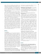
After targeted NGS with a lymphoma-dedicated panel, somatic missense and nonsense mutations were identified in 18 out of 30 PBL cases (60%). EBV-negative cases tend- ed to show a higher rate of mutations, as compared to EBV-positive cases (87.5% vs. 54%, respectively; c2 test, P>0.05) (Figure 1).
Interestingly the pattern of mutations was also different
haematologica | 2021; 106(4)
1121
