
Page 137 - 2021_04-Haematologica-web
P. 137
A novel flow cytometric panel to distinguish reactive and neoplastic PDC
ABC
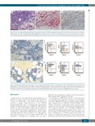
Figure 5. A case of reactive plasmacytoid dendritic cell proliferation positive for CD56 by immunohistochemistry. (A) A skin biopsy shows small clusters of plasma- cytoid dendritic cells (PDC), some with plasmacytoid morphology in a self-limited skin lesion, likely caused by an insect bite. The insert shows the low-power view of the skin biopsy. (B) Double staining for CD123/TCF4 highlights scattered and loosely clustered PDC. (C) CD56 immunostaining shows that many PDC are positive.
AC
B
D
Figure 6. Immunostain and flow cytometric analysis of a case of blastic plasmacytoid dendritic cell neoplasm before and after transplantation. (A, B) Immunostain using a dual-color TCF4/CD123 double stain showed scattered plasmacytoid dendritic cells (PDC) in both samples, before (A) and after (B) transplantation. (C, D) Flow cytometric analysis showed that a subset of PDC (red) in the pre-transplant sample (C) was aberrant (decreased CD38, negative CD2, decreased CD303 expres- sion) whereas all PDC in the post-transplant sample (D) showed a normal immunophenotype. CD56+ reactive PDC are highlighted blue in (C) and (D).
Discussion
In this study, we investigated the immunophenotype of BPDCN in a large cohort of 39 patients and compared it to that of reactive PDC. This study is the first to go beyond a simple characterization of the BPDCN immunophenotype, but to understand the immunophe- notypic aberrancy/alterations of BPDCN. Of particular interest, we show that CD56 is expressed in a small sub- set of normal/reactive PDC and therefore, CD56 alone is insufficient to differentiate BPDCN from reactive PDC, especially when the tumor burden is low. Through fur- ther characterization of these CD56+ normal PDC, we identified a combination of markers that can detect
BPDCN and distinguish neoplastic from non-neoplastic PDC in BM with a sensitivity of 0.01%.
The diagnosis of BPDCN at the time of initial presenta- tion, typically with a high tumor burden, is often straight- forward as BPDCN cells show a distinct immunopheno- type, being positive for HLA-DR, CD123 (bright), CD4, CD56, and absence of myeloperoxidase and monocytic markers as well as B- and T-cell lineage-defining markers. The neoplastic infiltrate can be further confirmed by immunohistochemical studies using CD123, TCL1 or a more specific TCF4/CD123 double stain. Basophils often have a similar level of CD123 expression but they are negative for HLA-DR. Monocytes, some hematopoietic precursors and acute myeloid leukemia blasts are positive
haematologica | 2021; 106(4)
1053
