
Page 136 - 2021_04-Haematologica-web
P. 136
W. Wang et al.
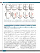
A
B
Figure 4. Representative cases of reactive and neoplastic CD56+ plasmacytoid dendritic cells. Gray: CD56– reactive plasmacytoid dendritic cells (PDC); blue: CD56+ reactive PDC; red: CD56+ neoplastic PDC. (A) CD56+ reactive PDC are consistently positive for CD2 and CD303, negative for CD7. CD38 expression is bright. (B) In contrast, neoplastic CD56+ neoplastic PDC are often negative for CD2 with decreased to negative CD303 expression. CD7 expression is often positive and CD38 expression level is often decreased. Focusing on CD56– PDC (gray) in both panels (A) and (B), these cells are positive for CD303 and CD38. CD2 shows a bimodal pattern of expression (both negative and positive cells present). A small subset of reactive PDC is CD7+ and these CD7+ reactive PDC are negative for CD56.
Table 2. The major immunophenotypic differences between CD56-positive reactive plasmacytoid dendritic cells and blastic plasmacytoid dendritic cell neoplasm.
CD2 positive
CD56-positive reactive PDC 100%
BPDCN 19%
CD7 positive
0%
64%
CD38 bright
100%
18%
CD303 positive
100%
44%
PDC: plasmacytoid dendritic cells; BPDCN: blastic plasmacytoid dendritic cell neoplasm.
CD4+CD64-CD56+HLA-DR+CD45dim+. Representative cases to illustrate our gating strategy are shown in Online Supplementary Figures S1 and S2. The sensitivity of this panel was validated to be 0.01% according to the MRD testing guideline from the College of American Pathologists (Online Supplementary Figure S3).
The ten-color MRD panel was tested prospectively in 19 BM samples from seven patients who had a confirmed diagnosis of BPDCN. These 19 samples included one for initial BM diagnosis and 18 samples for evaluation of resid- ual disease during the course of treatment. Using this flow cytometry panel, 12 (63%) samples were positive for BPDCN and the median number of aberrant cells was 0.05% of total nucleated cells (range, 0.008% to 56.5%). Of the 12 positive samples, one was detected as early relapse after stem cell transplant, with 0.01% of aberrant PDC. Of note, all samples had mixed reactive PDC in the background, serving as an internal comparison. All positive cases showed a similar immunophenotype to that identi- fied in the original diagnostic specimen and no significant immunophenotypic shift was observed. For patients who received anti-CD123 targeted therapy, CD123 expression was still maintained in BPDCN as well as normal PDC.
Flow cytometry versus immunohistochemistry in the assessment of minimal residual disease
We compared flow cytometry immunophenotyping and dual-color immunohistochemistry for TCF4/CD123 to
determine the relative performance of these assays in BM evaluation in the context of BPDCN after therapy. To achieve this, we first systematically assessed the number, distribution, and morphological characteristics of TCF4/CD123 dual-positive cells in 18 BM samples from patients without BPDCN. In such cases, PDC were few and often scattered, with a broad range of morphological char- acteristics that ranged from mature plasmacytoid forms to others with increased nucleus-to-cytoplasm ratio and occa- sional nuclear membrane convolutions. Although occasion- al loose PDC aggregates were identified, none of the cases had tight PDC aggregates or sheets of PDC. Next, we per- formed TCF4/CD123 double-stain immunohistochemistry in 14 cases with a history of BPDCN who had been evalu- ated for residual disease by flow cytometry immunopheno- typing. In these cases, TCF4/CD123 highlighted scattered PDC but could not reliably distinguish reactive from neo- plastic PDC. As shown in Figure 5, the TCF4/CD123 immunostain highlighted scattered PDC in a case of BPDCN prior to (Figure 6A) and after stem cell transplant in remission (Figure 6B), in both cases accounting for around 1-2% of the total cells in the BM. It is uncertain from the TCF4/CD123 immunostain whether these are aberrant or not. Flow cytometry, on the other hand, was capable of dif- ferentiating them: it detected neoplastic PDC mixed with normal PDC in the pre-transplant specimen (Figure 6C), whereas only reactive PDC but no aberrant PDC were detected in the post-transplant specimen (Figure 6D).
1052
haematologica | 2021; 106(4)
