
Page 135 - 2021_04-Haematologica-web
P. 135
A novel flow cytometric panel to distinguish reactive and neoplastic PDC
AB
CD
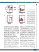
Figure 3. Differential immunophenotypic characteristics of blastic plasmacytoid den- dritic cell neoplasm and reactive plasmacy- toid dendritic cells. Blastic plasmacytoid dendritic cell neoplasm (BPDCN) cells often show increased HLA-DR, decreased CD123, decreased CD303, decreased CD38, and positive CD56 expression. Pink: basophils; blue: reactive plasmacytoid dendritic cells (PDC); red, neoplastic PDC; and gray: mono- cytes. (A) Both basophils and PDC are bright for CD123. Basophils are negative whereas PDC are positive for HLA-DR. In comparison to reactive PDC (blue), neoplastic PDC (red) often show decreased CD123 and increased HLA-DR expression. Monocytes (gray) are also positive for CD123 and HLA- DR, but their CD123 level is much lower than that of PDC. (B) Neoplastic PDC are positive for CD56 and negative for CD303. CD303 is positive in reactive PDC. (C) Neoplastic PDC often show decreased CD38 expression when compared to reac- tive PDC. (D) Reactive PDC are positive for CD33, and approximately half of BPDCN cases are negative for CD33.
and only one (6% in total) had a normal level of CD303 (Figure 3B). In contrast to bright CD38 expression in reac- tive PDC, CD38 expression was frequently downregulated in BPDCN cells, being decreased in 24 of 34 (70%) and negative in four of 34 (12%) (Figures 1 and 3C). While CD33 expression was positive in all cases of reactive PDC, it was only positive in 48% (16/33) of BPDCN cases.
We next focused on the difference between BPDCN and reactive CD56+ PDC. Unlike CD56+ reactive PDC that were uniformly positive for CD2, bright for CD38 and consistently negative for CD7, BPDCN cells were fre- quently negative for CD2 (81%), positive for CD7 (64%) and with decreased or negative (82%) expression of CD38 (Figure 4B) (Table 2). In contrast to the 100% positivity of reactive PDC for CD303, only 44% of BPDCN cases were positive. Using a combination of markers (CD2, CD7, CD56, CD303, CD38), none of the 39 BPDCN cases showed an immunophenotype exactly the same as that of CD56+ reactive PDC, which were CD56+/CD2+/CD7- /CD303+/CD38+bright.
Establishment and validation of a flow cytometry assay for minimal residual disease
Based on these findings, a one-tube, ten-color assay CD2/CD7/CD38/CD303/CD123/HLA- DR/CD64/CD4/CD45/CD56 was constructed (panel #3, Table 1). Detailed information, including the antibody clones and the fluorochrome attached to each antibody, is listed in Online Supplementary Table S2. CD123, HLA-DR, CD45, and CD64 were included to identify PDC that were CD123bright/HLA-DR+/CD64-/CD45dim+. In patients who received targeted therapy to CD123, an alternative gating strategy was also used to examine PDC that were
an expanded panel of markers, and demonstrated a remarkably consistent pattern. They were positive for CD2 (100%), negative for CD7 (100%), and showed bright CD38 (100%) expression in all 22 cases tested (Table 2). CD303 expression was also positive in all cases (100%), uniform in 13 (59%) and partial in nine (41%). A represen- tative case of CD56+ reactive PDC is shown in Figure 4A.
Of note, the expression of CD56 in normal/reactive PDC is not limited to BM samples. We analyzed a reactive PDC proliferation using immunohistochemistry in a patient who had a self-limited skin lesion, likely an insect bite, which had CD56 expression and had presented a diagnos- tic challenge at initial encounter (Figure 5).
Differential immunophenotypic characteristics of blastic plasmacytoid dendritic cell neoplasm and reactive plasmacytoid dendritic cells
The immunophenotype of BPDCN was compared to that of reactive PDC. In addition to being “positive” and “negative”, the markers of expression were also scored as “increased” or “decreased/partial” if the intensity difference was greater than one-third on a log scale (Figure 1). This comparison was facilitated by the presence of reactive PDC in some cases of BPDCN at initial diagnosis and in manycasesofBPDCNfollowingtherapy.Comparedwith reactive PDC, BPDCN cells showed brighter HLA-DR expression in 25 of 36 (69%) cases (Figure 3A), and lower CD123 expression in 28 of 36 (78%) cases (Figure 3A). In the latter cases, although decreased, CD123 levels in BPDCN were still higher than those of monocytes (Figure 3A). CD303, a marker that is consistently positive in nor- mal/reactive PDC, was only positive in seven of 16 (44%) BPDCN cases, of which six showed decreased expression
haematologica | 2021; 106(4)
1051
