
Page 79 - 2021_03-Haematologica-web
P. 79
Immunoprofiling and survival in DLBCL
AB
CD
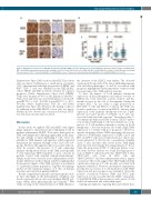
Figure 4. Membranous expression of HLA-ABC and β2 microglobulin (B2M) correlates with increased T-cell infiltration. (A) Representative images from B2M, HLA- ABC, and HLA-DR immunohistochemical (IHC) stainings. Scale bar 10 mm. (B) Results of the B2M, HLA-ABC, and HLA-DR IHC scoring. (C and D) Box plots visualizing the association of CD3+ T cells with HLA-ABC (C) and B2M (D) scores. P-values were determined by Kruskall-Wallis H (C) and Mann-Whitney U tests (D).
Supplementary Figure S8B), but not in the NLG Trial cohort (data not shown). Furthermore, a small group of patients (13%) characterized with high proportions of OX40+ and Ki67+ CD8+ T cells was identified in the HEL-DLBCL cohort. OX40 and Ki67 positivity trended for superior prognosis (Online Supplementary Figure S8C). FOXP3+ Tregs were frequent in 30% of the patients in the NLG- Trial cohort and were associated with inferior outcome (5- year OS 70% vs. 94%, P=0.041; 5-year PFS 71% vs. 91%, P=0.101) (Online Supplementary Table S8 and Online Supplementary Figure S9). However, the finding could not be validated in the HEL-DLBCL cohort (data not shown). Neither did we observe any correlation between TBET+ Th1-cells and outcome (data not shown).
Discussion
In this study, we applied GEP and mIHC with digital image analysis to characterize tumor infiltrating T cells in patients with primary DLBCL. We found a heterogeneous TME with lymphomas differing significantly in the num- ber and subtype of tumor-infiltrating T cells. First, we identified a TME immune cell signature, which separated the patients into immune cell high or “hot” and “cold” sub- groups. However, the signature did not associate with sur- vival. In contrast, the presence of T cells expressing immune checkpoint molecules in the TME, and especially T cells expressing TIM3 translated to poor outcome in patients treated with standard immunochemotherapy. Similar results were observed in two independent cohorts despite differences in their clinical variables, implying that the prognostic impact of the TME cytotypes is not limited to a particular patient population. Together, the findings underscore the regulatory impact of T cells on therapy resistance and survival in primary DLBCL.
Recent comprehensive genome and transcriptome stud- ies have shed light on the heterogeneity of DLBCL, and based on genomic drivers, uncovered multiple DLBCL sub- types, which differ phenotypically and clinically.6-8 Our study, focusing on the characterization of TME, illustrates
the diversity of the DLBCL even further. The observed variations in the amounts of the tumor-infiltrating immune cells and their phenotypes, as well as association with prognosis, highlight the clinical importance of the crosstalk between tumor cells, TME and host response.
To date, the impact of T-cell immune checkpoint expression on the pathogenesis and clinical outcome of DLBCL has been relatively scarcely studied, and has mainly focused on the role of the immune checkpoint receptor PD1.30 In one study, a high proportion of PD1+CD8+ T cells and PD-L1+ T cells in the TME was found to predict poor survival in DLBCL, whereas high expression of the immune checkpoint cytotoxic T-lym- phocyte-associated protein 4 (CTLA4) on T cells was associated with favorable outcome.14 In another study, T- cell immunoglobulin and ITIM domain (TIGIT) expres- sion in tumor infiltrating T cells was identified as a sup- pressor of T-cell mediated antitumor activity in B-cell lymphomas.31 A recent study has also found a high TIM3 expression to correlate with poor prognosis.32 However, specific subtyping of these TIM3-expressing cells was not addressed. Additionally, expression of TIM3, LAG3 and PD1 on CD8+ T cells was recently shown not only to rep- resent exhausted T cells but also a population of highly active T cells, and that other factors in the TME might affect the cells in eventually becoming exhausted.33 In our study, the proportion of PD1+ T cells alone did not corre- late with survival. Instead, we found that high propor- tions of TIM3+ T cells from all T cells, TIM3+CD4+ T cells, TIM3+CD4+ cells from all CD4+ cells and TIM3+CD4+CD3– cells have independent adverse impact on survival. The results suggest that TIM3 might identify a particular subgroup of T cells possibly associated with immune exhaustion. However, mechanistic experiments proving the association of T-cell dysfunction with TIM3 expression and poor prognosis are warranted. Given the positive correlation between TIM3 and IFNG gene expression, it is also possible that the TIM3+ cell popula- tion in DLBCL TME is heterogeneous and consists of cells with both exhausted and active phenotypes.
Checkpoint inhibitors targeting PD1 and CTLA4 have
haematologica | 2021; 106(3)
725
