
Page 68 - 2021_03-Haematologica-web
P. 68
M. Montesinos-Rongen et al.
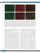
Figure 4. Recognition of SNRP by recombinant antibodies (recAb) corresponding to naïve B-cell receptors (nBCR) and tumor cell receptors (tBCR). (A-C) In primary lymphoma of the central nervous system (PCNSL) patient #17, the nBCR (FITC, A) derived from PCNSL patient #09, which did not react with SNRPC on the ProtoArray (Online Supplementary Table S4), shows only a weak staining of single tumor cells (arrows, A). Prominent expression of SNRPC in the nuclei of the majority of tumor cells in PCNSL patient #17 is evidenced by application of the commercial anti-SNRPC antibody (Cy3, B). The overlay (C) shows that there is no co-localization of both antibodies (arrows, C). Insert: high-power magnification of (C) x1000. *Erythrocyte autofluorescence. (D-F) tBCR (FITC, D) derived from PCNSL patient #09, which reacted with SNRPC on the ProtoArray (Online Supplementary Table S4), co-localizes with the commercial anti-SNRPC antibody (Cy3, E and F) that depicts prominent SNRPC expression by the tumor cells of PCNSL patient #17 (E). Arrows and arrowheads in (D) indicate prominent and moderate immunoreactivity of the tBCR, respec- tively, yielding a yellow (arrows, F) or orange (arrowheads, F) signal in the tumor cell nuclei in the overlay (F). Insert: high-power magnification of F, x1000. *Erythrocyte autofluorescence. Double immunofluorescence using rabbit anti-SNRPC (clone EPR16034, Abcam, Cy3) with nBCR (A) and tBCR (D) labeled with Mix’n Stain 488 Labeling Kit (Sigma). Microphotographs were taken with an Axiophot (Zeiss) and Zen 2 software (Zeiss). Original magnification x400 (objective: x40). Overlay of the microphotographs (C and F) and adjustment for contrast and brightness were performed with Adobe Photoshop software version CC. Similar results were obtained for staining with recAb derived from PCNSL patients #03 and #09, as well as for staining of sections derived from PCNSL patients #14, #21, and #24.
polyreactive BCR of the naïve precursor B cells. Comparison of the protein recognition pattern of nBCR and tBCR reveals increased polyreactivity of the GC-expe- rienced B cell with a particularly increased reactivity with proteins expressed in the CNS.
Gain of self- and polyreactivity during SHM was sup- ported by several experiments, i.e., reactivity with self- antigens in immunofluorescence with HEp-2 cells, ANA/ANCA ELISA at high, but not low BCR concentra- tion, ProtoArray analysis, immunoprecipitation, and topo- graphical immunohistochemistry. Comparability between the various methods is hampered by technical limitations due to specific characteristics of the respective analyses, e.g., different isoforms of proteins expressed may be pres- ent in the human brain and on the ProtoArray, a lack of knowledge of post-translational protein modifications and other metabolic modifications in the human brain. It is of note that our recAbs were expressed as IgG, a well-estab- lished technique frequently used by many groups.16,17 Recent studies demonstrated that the in vitro binding of at least certain polyreactive Igs differed from the IgM-BCR counterpart,18 that the C region may change the fine anti- body specificity for particular antigens,19 and that the iso- type shapes epitope specificity, antibody affinity, and functional activities.20 Thus, one cannot exclude with cer- tainty that some specificities may be confined to the solu- ble IgG antibody. However, it is unlikely that this holds true for all of the many specificities of all recAbs of our series. Moreover, the fact that the IgG generated from the tBCR did not show the same autoreactivity as their corre-
sponding IgG generated from the nBCR, further supports the concept that the expression of an IgM BCR as a solu- ble IgG antibody does not systematically lead to a gain of autoreactivity and hence serves as a kind of internal con- trol. N- and P-nucleotides of the CDR3 sequence cannot be reverted due to lack of corresponding germline sequences. However, it is unlikely that mutations persist- ing in CDR3, which can also modify antibody affinity and specificity, may affect the overall results of the entire PCNSL series, in particular, as PCNSL are characterized by unrestricted CDR3 length and amino acid composition and by an absence of stereotyped BCR sequences.4 Nevertheless, despite these technical limitations, all analy- ses indicated increased poly-/autoreactivity of the tBCR and a faulty GC reaction, thus extending previous data on polyreactive BCR of PCNSL and an involvement of numerous antigens in tumor cell selection.4,10,21
These observations fit into the concept that antigen selection plays an important role in driving BCR activity in PCNSL. There is growing evidence to suggest that BCR signaling in response to self and/or foreign antigens pres- ent in the target organ microenvironment sustains BCR signaling in various mature B-cell lymphoma entities including PCNSL, follicular lymphoma, splenic marginal zone lymphoma, mucosa-associated lymphoid tissue lym- phoma of the eye, and chronic lymphocytic leukemia (CLL).10,12,16,22,23 In CLL, B cells with unmutated BCR express highly polyreactive antibodies whereas most mutated CLL B cells do not.12 Mutated non-autoreactive CLL antibody sequences reverted into their germline
714
haematologica | 2021; 106(3)
ABC
DEF
