
Page 67 - 2021_03-Haematologica-web
P. 67
Impact of GC reaction on self-/polyreactivity in PCNSL
A
BC
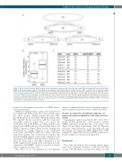
Figure 3. Tumor cell B-cell receptors (tBCR) recognize increased numbers of proteins in the central nervous system (CNS) as compared to naïve B-cell receptors (nBCR) in immunoprecipitation studies. (A) Quantitative Venn diagrams of immunoprecipitation analysis show the number of proteins that co-immunoprecipitate with at least one recombinant antibody (recAb) (upper panel). The middle panel shows numbers of proteins co-immunoprecipitating exclusively with nBCR or tBCR. The bottom panel depicts numbers of proteins co-immunoprecipitating nBCR or tBCR derived from either IGHV3+ or IGHV4-34+ primary lymphoma of the central nerv- ous system (PCNSL). Proteins co-immunoprecipitating with both recAb are shown in the intersection. (B) BoxPlot diagram of immunoprecipitation shows the numbers of proteins co-immunoprecipitating with at least one recAb. Only proteins co-immunoprecipitating with either nBCR or tBCR were analyzed. Statistical significance was determined by exact Wilcoxon signed rank test. (C) Numbers of proteins co-immunoprecipitating with nBCR and tBCR shown for individual PCNSL cases. In addi- tion, numbers of shared target proteins are depicted.
method, double immunofluorescence of SNRPC expres- sion was performed.
SNRPC was differentially recognized by recAb derived from nBCR and tBCR on the ProtoArray (nBCR: nega- tive, tBCR: positive, PCNSL patients #01, #03, #09). RecAB derived from the nBCR of PCNSL patients #01, #03, and #09 did not co-localize with the commercial anti-SNRPC antibody (Figure 4A-C). In contrast, the recAb derived from the tBCR co-localized with the com- mercial anti-SNRPC antibody demonstrating tumor cell SNRPC expression (Figure 4D-F). RecAb staining was remarkably less strong compared to the commercial monoclonal anti-SNRPC antibody, further indicating their lower affinity. This observation was further sup- ported by ELISA analysis of the recAb compared to the commercial anti-SNRPC antibody (Online Supplementary Figure S8). Here, only a very high concentration of the recAb together with a much lower concentration of the commercial anti-SNRPC antibody revealed reactivity (Online Supplementary Figure S8).
Thus, these in situ topographical proof-of-principle
analyses confirmed the ProtoArray data further support- ing low-affinity and polyreactivity of nBCR and tBCR.
Proteins recognized by recombinant antibodies are expressed in primary lymphoma of the central nervous system
Topographical analysis revealed that 90% (18 of 20) of PCNSL (PCNSL patients #11 - #30). showed a nuclear SNRPC expression in the majority of the tumor cells (Figure 5A and Online Supplementary Table S7). Neurons, astrocytes, oligodendrocytes, a few microglial cells and macrophages also expressed SNRPC (Figure 5B-F). GEP15 confirmed a significantly increased SNRPC expression in PCNSL compared to normal brain (Figure 5G).
Discussion
This study demonstrates that reverting somatic muta- tions of the V-derived sequences of the VH and VK/L domain of PCNSL tBCR can yield a low-affinity, self- and
haematologica | 2021; 106(3)
713
