
Page 53 - 2021_03-Haematologica-web
P. 53
Aberrant expression of SCF in CLL
ABC
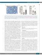
Figure 5. Functional implications of stem cell factor in chronic lymphocytic leukemia cells. (A) Immunohistochemical staining showing weak nuclear immunoposi- tivity for c-Kit of the scarce chronic lymphocytic leukemia (CLL) cells residing in the lymph nodes. (B) Real-time quantitative polymerase chain reaction analysis of KIT transcripts in stem cell factor (SCF)high CLL cases and SCFlow CLL cases. (C) Mitochondrial mass in CLL cells treated or not (control) with 100 ng/mL SCF for 48 h. Interconnected dots represent one case in two different conditions and bars represent median values. The Wilcoxon P test was applied to evaluate statistical signif- icance; *P<0.05, **P<0.01, ***P<0.001. FD: fold difference.
controlled by mitochondrial ROS.35 Additionally, SCF is amplified in CLL cells in parallel with HIF-1a during long term co-cultures with the mesenchymal stroma, likely as a reflection of the upregulation of glycolytic markers (e.g., c-myc).35,36,47 Moreover, proliferation centers, known to be hypoxic in CLL, were also found to be highly SCF-posi- tive.48 For these reasons, the herein identified association of HIF-1a with SCF cannot be viewed as paradoxical and recalls findings in certain solid malignancies such as, pan- creatic ductal adenocarcinoma, in which HIF-1a can func- tion as a transcription factor for the KITLG gene.44
The convergence of immunoproliferative and redox sig- naling for the regulation of SCF in CLL is further support- ed by the results of our studies with ibrutinib, a BTK inhibitor that: (i) impinges on proliferative signals con- veyed to CLL cells by their neighboring cells via several receptors, such as BcR, TLR and CD40; (ii) suppresses the expression of several chemoattractants and inflammatory cytokines from CLL cells; and, (iii) dishevels the metabo- lism of CLL cells, inciting perturbations in their mito- chondrial dynamics.38,39 Indeed, longitudinal profiling revealed significant mitochondrial changes in CLL cells after treatment, along with significant suppression of SCF and HIF-1a, thus underlining the importance of immune signaling, hypoxia and mitochondrial ROS for the overex- pression of SCF in CLL.
Our study documented the presence of low-abundant KIT transcripts in CLL cells while also showing that their stimulation with soluble SCF suppressed mitochondrial mass, indicative of functional SCF/c-kit signaling. Besides a possible SCF/c-kit autocrine interaction within CLL cells, SCF may also function as a receptor with signaling capac- ity, as was recently revealed in thymic cells and could arguably apply to CLL cells as well.49 The detection of large numbers of c-kit+ mast cells, especially in CLL bone marrow biopsies, highlights paracrine stimulation of c-kit reactive microenvironment by SCF+ CLL cells as an alter- native, not mutually exclusive mode for SCF-induced modulation of CLL cell physiology, which merits thor- ough investigation in future studies.
In conclusion, the present study offers novel perspec- tives on microenvironmental interactions in the CLL
microenvironment by focusing on the previously unex- plored role of SCF. On the basis of our findings, SCF emerges as an important player in the regulation of redox and proliferative homeostasis in CLL cells.40 Considering our finding that SCF is associated with adverse prognosis and modulated by the CLL microenvironment, selective targeting of SCF may represent a novel therapeutic para- digm for CLL.
Disclosures
KS has received research support and honoraria from Janssen Pharmaceutica, Gilead Sciences and Abbvie SA.
Contributions
GIG and SN designed the research, performed the study and wrote the manuscript; NP designed the research and assisted in data analysis, and interpretation; KK and EC assisted in research, data analysis, and interpretation; TK provided samples and performed the immunohistochemical analysis; TM performed data and statistical analysis and assisted in data interpretation; NS and AA provided patients’ samples; EP assisted in data interpretation and revised the manuscript; AST and KS designed and supervised the research and wrote the manuscript.
Funding
This work was supported in part by Greek Government Departmental funds for graduate student research distributed to
the Laboratory of Pharmacology (to AST) of the Department of Pharmacy of Aristotle University of Thessaloniki, Greece; the GCH-CLL project funded by ERA NET TRANSCAN-2 Joint Translational Call for Proposals 2014 (JTC 2014); the ODYSSEUS Program, implemented under the “Action for the Strategic Development on the Research and Technological Sector”, funded by the Operational Program "Competitiveness, Entrepreneurship and Innovation" (NSRF 2014-2020) and co- financed by Greece and the European Union (European Regional Development Fund); the NEoteRIC, H2020-ICT-2018- 20/H2020-ICT-2019-2 (grant agreement number: 871330). Part of this work will be submitted to the faculty of the Department of Pharmacy of Aristotle University of Thessaloniki, Greece as part of the requirements for awarding a doctorate degree in Pharmacology to GIG.
haematologica | 2021; 106(3)
699
