
Page 52 - 2021_03-Haematologica-web
P. 52
G.I. Gavriilidis et al.
measured mitochondrial dynamics. We found that SCF significantly suppressed mitochondrial mass (n=9, FD:1.2; P=0.0039) (Figure 5C, Online Supplementary Figure S7D), indicating functional SCF/c-kit signaling in CLL cells. There was no difference in the anti-oxidant effect of SCF between SCFhigh and SCFlow cases.
Discussion
In the present study, we assessed SCF expression in a well-characterized CLL cohort and also explored how the microenvironment might modulate SCF protein levels in CLL cells. We found that SCF is overexpressed in CLL B cells compared to healthy B cells and that the membrane isoform (KL-2) predominates. Interestingly, a striking dis- crepancy was noted between elevated SCF protein levels versus low-abundant KITLG transcripts, indicative of post- translational modifications or distinct decay kinetics for KITLG transcripts and SCF protein levels which merit fur- ther molecular investigation.
By correlating SCF expression with the clinicobiological characteristics of the CLL patients included in the study, we report for the first time a strong association between SCF overexpression and adverse prognosis in CLL since increased SCF protein levels were associated with several adverse prognostic biological features (e.g., CD38 and ZAP70 positivity, unmutated IGHV genes) as well as shorter overall survival and TTFT. These findings are in line with a growing body of evidence regarding the nega- tive prognostic significance of SCF in solid tumors e.g., hepatocellular carcinoma, colorectal carcinoma, prostate cancer and pancreatic ductal adenocarcinoma.42-45 Moreover, a significant association was identified between SCF overexpression and trisomy 12, which might be attributed to a gene dosage effect considering that the KITLG gene locus is located on chromosome 12.12
Stimulation of immune receptors (BcR, TLR9, CD40) was found to modulate the levels of both membrane and soluble SCF, thus highlighting the dynamic nature of SCF expression in CLL. It is worth mentioning that strong SCF protein expression characterized the rapidly dividing, Ki- 67+ CLL cells, while SCF downmodulation using siRNA decreased the proliferation of MEC1 CLL cells. These findings are in keeping with the herein presented immunohistochemical evidence of intense expression of SCF in the bone marrow and lymph nodes of CLL patients, where the proliferating fraction of CLL cells resides.9
Our work also suggests that SCF protein levels are affected by mitochondrial redox homeostasis of CLL cells within the tumor microenvironment. Such modulation of SCF expression by oxidative stress has been previously reported in astrocytes and endothelial cells, prompting the hypothesis that in the short-term SCF contributes to com- pensatory cellular repair mechanisms while in the long- term it can further incite the inflammatory milieu by attracting immune cells.18,19 Recent studies in healthy B cells, T cells and macrophages showed that inflammatory responses to immune signals are wired by mitochondrial dynamics.46 In the case of CLL cells, mitochondrial ROS levels are significantly higher than in healthy B cells due to intensified mitochondrial biogenesis. This aberrant oxida- tive stress affects CLL cells and their surrounding cells by establishing inflammation and immune evasion in the
CLL milieu.30 Our data point towards a similar trend for SCF regulation in CLL by showing for the first time that proliferating CLL cells significantly augment mitochondri- al ROS in tandem with SCF protein levels.
A possible causal relationship between mitochondrial ROS and SCF was suggested by our finding that both were significantly suppressed in short-term co-culture with HS-5 mesenchymal stromal cells. In this context, the mesenchymal stroma was recently found to supply CLL cells with cysteine, thus leading to a marked increase of glutathione, alluding to the anti-oxidant capacity of mes- enchymal stromal cells in the CLL microenvironment.31 Unsurprisingly, therefore, the suppression of SCF in CLL cells by HS-5 mesenchymal stromal cells was reversed once the oxidative stimuli of H2O2 and CpG/CD40L were introduced in the co-cultures.
Our study also revealed a significant correlation between the protein levels of SCF and HIF-1a in CLL cells. Recent experiments with the mitochondrial complex I inhibitor BAY 87-2243 in CLL cells have shown that, as in several other solid tumors, the stability of HIF-1a is tightly
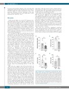
AB
CD
Figure 4. Ibrutinib leads to downregulation of mitochondrial mass, hypoxia-
inducible factor-1a and stem cell factor. (A) Mitochondrial mass in chronic lym-
phocytic leukemia (CLL) samples before and after 1 month of ibrutinib therapy
determined by flow cytometry analysis for viable CLL cells. (B) Hypoxia-inducible
factor (HIF)-1a protein expression in CLL samples before and after 1 month of +
ibrutinib therapy determined by flow cytometry analysis for viable HIF-1a CLL
cells. (C) Stem cell factor (SCF) protein expression in CLL samples before and
after 1 month of ibrutinib therapy determined by flow cytometry for viable SCF+
CLL cells. (D) SCF protein expression (determined by flow cytometry for viable
+
SCF CLL cells) in samples from CLL patients after 1 month of ibrutinib therapy
and loss of response to treatmment. Interconnected dots represent one case in two different conditions and bars represent median values. The Wilcoxon P test was applied to evaluate statistical significance. *P<0.05, **P<0.01. MFI: mean fluorescence intensity; ibr: ibrutinib
698
haematologica | 2021; 106(3)
