
Page 269 - 2021_03-Haematologica-web
P. 269
Letters to the Editor
that these were composed of 62,4% protoporyphin IX (PPIX) (5.43 mmol/L), 19.9% URO (1.73 mmol/L) and 15% COPRO (1.30 mmol/L). At month 23 of treatment, erythrocyte porphyrin chromatography showed a large decrease in URO (1.1%, 0.08 mmol/L; 95% decrease com- pared to baseline) and COPRO (8.6%, 0.63 mmol/L, 51% decrease compared to baseline) and an increase in the amount of PPIX (89.3%, 6.52 mmol/L; 20% increase; 76% of zinc PPIX) due to iron-deficiency anemia. Hemoglobin levels remained greater than 10 g/dL. Clinical tolerance was good with moderate asthenia. The patient’s urine became clear. No adverse effects were detected. The bio- logical and clinical improvement allowed the patient to have unrestricted exposure to sun during the summer without showing any signs of blistering.
The impact of iron metabolism on the clinical expres-
A
sion of CEP is reinforced by the observation of a consan- guineous Pakistani family with three of four children diagnosed with CEP (Figure 2A). Their UROS genotype was associated with a mild CEP phenotype in an Indian family.6 The proband of the Pakistani family (subject II1), a 9-year-old girl, exhibited photosensitivity, skin fragility and blistering, hirsutism and mild, compensated hemoly- sis without iron deficiency. She had markedly elevated urine and plasma porphyrin levels (Figure 2B). Her 5-year-old affected brother (subject II3) never experi- enced any symptoms of CEP despite unrestricted expo- sure to sun. His plasma porphyrin levels were in the nor- mal range. Urine porphyrin levels, mostly consisting of isomer I, were almost normal and 56-fold lower than those of his sister. He had mild aregenerative microcytic anemia without hemolysis due to a profound iron defi-
B
C
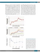
Figure 3. In vitro erythroid cell porphyrin analysis. Cells were harvested on day 18 for assessment of total erythroid cell porphyrins and porphyrin sepa- ration by high performance liquid chromatography with fluorescence detection. Erythrocyte, plasma and urine porphyrins were measured as previously described.15 (A) Porphyrin levels in erythroid cells derived from peripheral CD34+ cells, normalized by protein content after culture in increasing concen- trations of holo-transferrin (Holo-Tf). Porphyrins were not detectable in the 20 mg/mL condition, presumably because of the technique’s insufficient sensitivity. (B) Quantification and separation of the different types of porphyrins in erythroid cells by chromatography. (C) The ratio of protoporphyrin IX (PPIX) to coproporphyrin (Copro) level plus uropro- porphyrin level ranged from approximately 0.3 in iron-depleted conditions to 0.15 in iron-replete con- ditions.
haematologica | 2021; 106(3)
915
