
Page 267 - 2021_03-Haematologica-web
P. 267
Letters to the Editor
Phlebotomy as an efficient long-term treatment of
congenital erythropoietic porphyria
Congenital erythropoietic porphyria (CEP, MIM 263700) is a rare autosomal recessive disease caused by impaired activity of uroporphyrinogen III synthase (UROS), the fourth enzyme of the heme biosynthetic pathway.1 Accumulation of porphyrins in red blood cells, mainly uroporphyrinogen I (URO I) and copropor- phyrinogen I (COPRO I), leads to ineffective erythro- poiesis and chronic hemolysis. Porphyrin deposition in the skin is responsible for severe photosensitivity result- ing in bullous lesions and progressive photomutilation. Treatment options are scarce, relying mainly on support- ive measures and, for severe cases, on bone marrow transplantation (BMT). Increased activation of the heme biosynthetic pathway by gain-of-function mutations in ALAS2, the first and rate-limiting enzyme, results in a more severe phenotype.2 Iron deficiency promotes the binding of iron regulatory protein to the 5’ untranslated region iron-responsive elements of ALAS2 mRNA and therefore decreases its translation.3 Egan et al. observed a 32-year-old woman with CEP who exhibited sponta- neous improvement in photosensitivity and hemolysis
after chronic gastrointestinal bleeding that resulted in iron deficiency.4 They treated her with an iron chelator, resulting in correction of the hemolysis, decreased por- phyrin levels and improved quality of life with reduced photosensitivity. We recently identified three CEP sib- lings with phenotypes ranging from moderate to asymp- tomatic which were modulated by iron availability, high- lighting the importance of iron metabolism in the disease. Based on these data, we prospectively treated a CEP patient with phlebotomies to investigate the feasibility, safety and efficacy of this treatment. We observed dis- continuation of hemolysis and a marked decrease in plas- ma and urine porphyrins. The patient reported a major improvement in photosensitivity. Finally, erythroid cul- tures were performed, demonstrating the role of iron in the rate of porphyrin production.
The study was conducted in accordance with the World Medical Association Declaration of Helsinki ethi- cal principles for medical research involving human sub- jects.
A 49-year-old Caucasian female was treated for CEP by repeated phlebotomies for almost 2 years. She was diag- nosed with CEP at the age of 25 after experiencing symp- toms since the age of 4, including photosensitivity with skin fragility, blistering and scarring, mouth lesions with
A
B
C
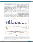
Figure 1. The patient’s clinical and biological parameters throughout treatment. (A) Urine porphyrins (gray), plasma porphyrins (blue) and ferritin (dark red). (B) Erythrocyte porphyrins (brown), protoporphyrins (light green), coproporphyrins (purple) and uroporphyrins (orange). (C) Hemoglobin (dark blue), reticulocytes (red) and haptoglobin (dark green). At baseline, the urine porphyrins/creatinine ratio ranged from 1845 to 2565 nmol/mmol (reference <30 nmol/mmol), plasma porphyrin concentrations ranged from 227 to 289 nmol/L (reference <20 nmol/L), reticulocyte counts ranged from 115 to 128 x 109/L (reference range, 20 - 80 x 109/L), the haptoglobin level was 0.16 g/L (reference range, 0.56 - 2 g/L) and ferritin ranged from 120 to 150 mg/L (reference range, 18 - 160 mg/L). Over the entire treatment course, ferritin levels were not greater than 17 mg/L.
haematologica | 2021; 106(3)
913
