
Page 268 - 2021_03-Haematologica-web
P. 268
914
Letters to the Editor
erythrodontia, microstomia and gum recession, sclero- dermiform retraction of the skin on the face and hands and loss of a distal phalange. She showed moderate chronic hemolysis. Genetic testing revealed a homozy- gous UROS variant, p.Gly58Arg, resulting in markedly decreased UROS activity in red blood cells (1.7 U/mg Hb/h, N>6). The patient was treated mainly with sup- portive measures, such as sun avoidance. At diagnosis, urine porphyrin levels were 2642 nmol/mmol of creati- nine (URO 1602 nmol/mmol and COPRO 1040 nmol/mmol). Treatment started with the removal of 100 mL of blood twice a month for 1 month. The volume of blood was then increased by 50 mL each month or each 6 weeks until reaching 300 mL. This gradual increase of the volume of blood withdrawn was to avoid a strong induction of erythropoiesis. The iron store was efficiently depleted 8 months after the first phlebotomy, and the fer- ritin level decreased to 10.4 mg/L (Figure 1A). Concomitantly, we observed a significant decrease in urine and plasma porphyrin levels to 389 nmol/mmol and 50 nmol/L, respectively, corresponding to 79% and 85% decreases compared to the baseline levels. A pro-
gressive normalization of haptoglobin levels occurred which was associated with a decrease in reticulocyte count, showing discontinuation of hemolysis (Figure 1C). Photosensitivity markedly improved, and the urine became clearer. Phlebotomies were discontinued for 3 months, resulting in an increase in urine and plasma por- phyrins (urine porphyrins 1956 nmol/mmol, plasma por- phyrins 219 nmol/L) associated with worsening photo- sensitivity. Phlebotomies were reintroduced at month 11, on a monthly basis, progressively increasing the blood volume over 7 months until reaching 300 mL. Phlebotomies were then continued at 200 mL monthly. At month 18, low levels of urine and plasma porphyrins were obtained again (460 nmol/mmol and 31 nmol/L, respectively). Finally, at month 23, urine and plasma por- phyrin levels were reduced to 271 nmol/mmol and 26 nmol/L (83% and 91% reductions compared to baseline), close to levels observed after BMT.5 During the course of treatment, total erythrocyte porphyrins levels were mod- erately reduced (8.7 mmol/L at baseline, 7.3 mmol/L at month 23; 16% decrease) (Figure 1B). Chromatography of erythrocyte porphyrins prior to phlebotomies revealed
A
B
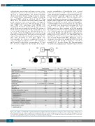
Figure 2. The pedigree of a family with congenital erythropoietic porphyria and biological parameters at diagnosis. (A) The family pedigree showing three of four siblings diagnosed with congenital erythropoietic porphyria (M: UROS c.660+4delA, +: wild-type allele). Genetic testing confirmed the diagnosis: patients II1, II3 and II4 are homozygous for the UROS mutation c.660+4delA, previously reported.6 (B) The siblings’ laboratory values at diagnosis. CRP: C-reactive pro- tein; UROS: uroporphyrinogen III synthase; Zn: zinc; ND: not determined. NA: not applicable.
haematologica | 2021; 106(3)
