
Page 154 - 2021_03-Haematologica-web
P. 154
A. Nai et al.
A
BC
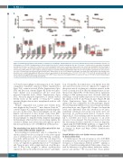
Figure 3. Hematologic parameters and analysis of erythropoiesis in wild-type and Ncoa4-ko mice fed an iron-deficient diet. Ncoa4-ko and wild-type (wt) mice (of both genders) on a Sv129/J background were fed an iron-deficient diet for 6 months starting from the age of 3 months. Complete blood counts were determined periodically. (A) Values are shown for red blood cell count, hemoglobin levels, mean corpuscular volume and mean corpuscular hemoglobin. (B) Percentage of Ter119+ cells on alive cells and subpopulation composition (determined as described in Figure 1B) both in the bone marrow and in the spleen of 9-month old iron- deficient wt and Ncoa4-ko mice. The complete statistical analysis is reported in Online Supplementary Table S5. (D) Colony numbers generated from 104 bone mar- row cells of 9-month old iron-deficient wt and Ncoa4-ko mice. Mean values of four or five animals per genotype are shown. Error bars indicate the standard error. Asterisks refer to statistically significant differences between age-matched wt and Ncoa4-ko mice. *P<0.05; **P<0.01; ***P<0.005. ID: iron-deficient; RBC: red blood cells; Hb: hemoglobin; MCV: mean corpuscular volume; MCH: mean corpuscular hemoglobin; BM: bone marrow, SP: spleen; CFU-GM: colony-forming unit-gran- ulocyte/monocyte; BFU-E: burst-forming unit-erythroid.
of Ncoa4-ko macrophages in releasing iron in vivo, despite comparable transferrin saturation (Online Supplementary Figure S5A), serum iron levels (Online Supplementary Figure S5B) and liver iron content (Figure 4E) in the two geno- types. Splenic iron accumulation did not result from increased hepcidin levels (Online Supplementary Figure S5C), but from impaired degradation of ferritin. Indeed ferritin levels in the spleen of Ncoa4-koko BM animals remained higher than in mice transplanted with wt cells (Figure 4F).
Despite comparable iron content, liver ferritin levels were higher in the Ncoa4-koko BM mice than in Ncoa4-kowt BM controls (Figure 4G), suggesting impaired ferritin degrada- tion also in this tissue. Since macrophages are the only hepatic cells derived, at least in part, from donor BM,21 these results further confirm the reduced ability of Ncoa4- ko macrophages to degrade ferritin. Limited macrophage ferritinophagy, which restricts iron recycling, explains the severity of anemia in iron deficiency and supports a preva- lent non-autonomous role of NCOA4 in erythropoiesis.
The circulating iron levels in Ncoa4-ko mice fail to rise upon acute erythropoietic expansion
Iron recycling is essential not only to compensate for chronic anemia but also in response to the acute expan- sion of erythropoiesis, such as after bleeding or stimula- tion with erythropoietin. To verify the role played by NCOA4 in response to an acute increase of iron demand in vivo, we exploited a published protocol,22,23 treating wt and Ncoa4-ko mice with a single injection of erythropoi- etin (8 IU/g body weight) to induce erythropoietic expansion, increased erythroferrone release24 and inhibi-
tion of hepcidin. In normal mice, iron uptake from the diet and release from stores are enhanced to supply ery- thropoietic needs, resulting in a transient increase in the levels of serum iron 15 h after the administration of ery- thropoietin22 (Figure 5A). At this time point no significant changes of BM erythropoiesis were observed, while the percentage of early erythroid precursors was increased in the spleen in both wt and Ncoa4-ko mice (Figure 5B and Online Supplementary Figure S6A). The induction of splenic Erfe was comparable in both genotypes (Figure 5C) and hepcidin inhibition was even stronger in mutant mice than in wt ones (Figure 5D). Despite low hepcidin levels and differently from the situation in wt mice, transferrin saturation and serum iron levels were not increased in erythropoietin-treated Ncoa4-ko mice (Figure 5E, F). These findings indicate that the latter mice fail to mobilize iron stores in response to an acute increase of iron demand. The hypoferremia observed in the latter animals likely contributes to decrease hepcidin, suppressing the BMP-SMAD pathway, as suggested by the concomitant reduction of Id1 mRNA, in the absence of changes in Bmp6 and Bmp2 (Online Supplementary Figure S6B-D).
Ncoa4 deletion does not further worsen anemia in thalassemic mice
To further prove the non-autonomous role of NCOA4 in erythropoiesis, we investigated the effect of Ncoa4 het- erozygous or homozygous deletion in Hbbth3/+ animals, a model of transfusion-independent thalassemia, character- ized by deficiency of β-globin chains, ineffective erythro- poiesis and anemia.14 We reasoned that Ncoa4 genetic
800
haematologica | 2021; 106(3)
