
Page 153 - 2021_03-Haematologica-web
P. 153
Macrophage ferritinophagy supports erythropoiesis
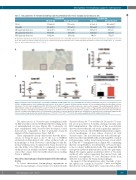
Table 1. Iron parameters of 9-month old wild-type and Ncoa4-knockout mice fed a standard and an iron-poor diet.
Standard diet
Iron-poor diet
Ncoa4-ko (n=5)
14.10±2.88**
74.5±13.4* 165.9±5.0 632±155 772±53
TS(%)
WT (n=5-10)
57.21±2.58
296.1±34.5 522.1±37.4 4897±713 1382±168
Ncoa4-ko (n=6-13) 57.60±1.80
331.9±55.6 596.6.5±37.5 3518±417 1276±122
WT (n=4)
36.70±5.03
176.9±24.5 136.6±10.9 1161±215 748±59
SI(mg/dL)
LIC (mg iron/g dry tissue) SIC (mg iron/g dry tissue) KIC (mg iron/g dry tissue)
TS: transferrin daturation; SI: serum iron; LIC: liver iron content; SIC: spleen iron content; KIC: kidney iron content. The values reported are the mean ± standard error for each group.The number of animals analyzed in each group is indicated in the header.Asterisks refer to statistically significant differences between Ncoa4-knockout (ko) and wild- type (wt) mice fed the same diet. *P<0.05; **P<0.01.
AB
CDE
Figure 2. Analysis of the iron phenotype of 9-month old wild-type and Ncoa4-ko mice fed a standard diet. Ncoa4-ko and wild-type (wt) mice (of both genders) on a Sv129/J background were fed a standard diet until sacrifice at the age of 9 months. (A) Representative pictures of Perls staining performed on duodenal sections (thickness of section 5 μm; magnification 20X, 40X in the inset). (B-D) Quantitative real-time polymerase chain reaction analysis of hepcidin (Hamp) (B), inhibitor of differentiation 1 (Id1) (C) and transferrin receptor 1 (Tfr1) (D) to measure mRNA levels in the liver relative to those of hypoxanthine phosphoribosyltransferase 1 (Hprt1). Data were normalized on a wild-type mean value of 1. (E) Western blot and relative densitometric analysis of ferritin H protein levels in the liver. Tubulin was used as a loading control; Mean values of 6-13 animals per genotype are shown. Error bars indicate the standard error. Asterisks refer to statistically significant dif- ferences between age-matched wt and Ncoa4-ko mice. *P<0.05; **P<0.01; ***P<0.005. RQ: relative quantification; FTH: ferritin H; Tub: tubulin.
The microcytosis of Ncoa4-ko mice transplanted with wt BM was fully corrected with their MCV reaching levels of untransplanted wt mice (Figure 4B). Conversely, wt mice transplanted with Ncoa4-ko BM developed a mild microcytosis, comparable to that of germ-line Ncoa4-ko animals (Online Supplementary Figure S4B-E).
These experiments clearly demonstrate that the micro- cytosis of Ncoa4-ko mice is due to the loss of NCOA4 exclusively in BM-derived cells, either erythroid cells or macrophages or both. However, considering not only the normal erythropoiesis documented in Ncoa4-ko mice but also the ability of ko BM cells to completely reconstitute erythropoiesis in lethally irradiated animals, our conclu- sion is that a major defect in erythroid precursors lacking Ncoa4 is unlikely.
Ncoa4-ko macrophages display impaired ferritinophagy in vivo
To better characterize ferritinophagy impairment in Ncoa4-ko macrophages, Ncoa4-ko mice reconstituted with
Ncoa4-ko (Ncoa4-koko BM) or wt BM cells (Ncoa4-kowt BM) were fed a low iron diet for 3 months starting 2 months after BM transplantation. The duration of this iron-deficient diet was shorter than the one used in germ-line animals to avoid the activation of the NCOA4-independent compen- satory mechanisms of iron release observed in total Ncoa4-ko mice. The iron-deficient diet reduced hemoglo- bin levels, MCV and MCH without affecting RBC count in both genotypes, with a trend toward a more severe effect in Ncoa4-koko BM transplanted mice (Figure 4B), confirming that the susceptibility to iron-deficiency of Ncoa4-ko mice is due to a defect in BM-derived cells. The comparable BM and spleen erythroid differentiation in the two genotypes (Figure 4C) argues against an intrinsic defect of Ncoa4-ko erythroid cells, rather suggesting that microcytosis and increased susceptibility to iron deficiency result from defective iron release by Ncoa4-ko macrophages.
In support of this interpretation, after 3 months of diet, the spleen iron content in Ncoa4-koko BM mice was higher than that in controls (Figure 4D), consistent with a defect
haematologica | 2021; 106(3)
799
