
Page 128 - 2021_03-Haematologica-web
P. 128
X. Li et al.
ABCD
EFG
HIJK
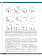
Figure 1. Reduced expression sHLA-G and mHLA-G in immune thrombocytopenia patient plasma. (A) The plasma concentration of secreted human leukocyte anti- gen-G (sHLA-G) was assayed by enzyme linked immunosorbent assay (ELISA). Immune thrombocytopenia (ITP) patients positive for anti-platelet autoantibodies (n=34) showed significantly reduced plasma sHLA-G compared to ITP plasma negative for anti-platelet autoantibody (n=16) and compared to healthy controls (n=15). (B) No significant differences in plasma sHLA-G were found between ITP patients with double positive (n=11), anti-GPIIb/IIIa positive (n=11), and anti-GPIb/IX positive (n=12) plasma. However, each of these groups showed reduced plasma HLA-G compared to double negative ITP plasma (n=16) and healthy controls (n=15). (C) sHLA-G level in plasma of patients with high platelets (≥10x109/L; n=19) was significantly higher than those with low platelets (<10x109/L; n=31). (D) Patients responded to high-dose dexamethasone (HD-DXM) treatments exhibited increased sHLA-G level (n=14). (E-G) Plasma concentration of sHLA-G correlated with platelet counts in autoantibody positive, negative patients and healthy controls. *P<0.05; **P<0.01. (H-K) Cell surface expression of membrane-bound HLA-G (mHLA-G) on CD4+, CD8+, CD14+, and CD19+ cells in ITP patients (n=17) and healthy controls (n=15).
effect on monocytic Fcg-receptor (CD16) expression. Therefore, no difference was observed in the antibody- mediated platelet phagocytic capability between rhHLA- G modulated and unmodulated groups (Online Supplementary Figure S3), indicating that rhHLA-G protects ITP platelets through mechanisms other than by inhibiting platelet phagocytosis.
rhHLA-G suppressed DC maturation and T-cell proliferation in vitro
In order to assess whether rhHLA-G inhibits maturation of monocyte-derived DC, surface expression of CD80 and CD86 on DC was analyzed by flow cytometry. DC from ITP patients expressed higher levels of CD80 and CD86 than those from healthy controls. In the presence of rhHLA-G, CD80 and CD86 expression was markedly downregulated in both ITP patients and healthy controls (Figure 5A-D).
DC maturation, which increases the expression of cos-
to induce platelet apoptosis. PBMC from ITP patients induced significantly higher apoptosis of both autologous and healthy control platelets compared with healthy con- trols and the addition of rhHLA-G attenuated platelet destruction by ITP PBMC. The decrease of platelet apop- tosis demonstrates a protective effect of rhHLA-G in ITP. In order to determine whether the platelet apoptosis was mediated by PBMC cytotoxicity and by the platelet them- selves, healthy control PBMC were cultured with autolo- gous platelets and ITP platelets respectively. The results showed that rhHLA-G modulation did not change the ability of healthy control PBMC to induce both autolo- gous and ITP platelet apoptosis (Figure 4).
Since Fcg-receptor-mediated platelet phagocytosis is one of the most important mechanisms for antibody-mediated platelet destruction in ITP, we detected the Fcg-receptor (CD16) expression and antibody-mediated platelet phago- cytic capability by monocytes/macrophages. Our results showed that treatment of rhHLA-G had no significant
774
haematologica | 2021; 106(3)
