
Page 127 - 2021_03-Haematologica-web
P. 127
HLA-G corrects dysfunction of immune cells in ITP
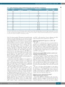
Table 2. Clinical characteristics of immune thrombocytopenia patients treated with high-dose dexamethasone.
Patient No.
37
38
39
Sex/Age Autoantibodies Bleeding symptoms
Platelet counts (x109/L) before after
7 78R
9 23NR 2 166R 3 56R 1 86R 2 98R
(yr) Anti-GPIb/IX
F/42 +
F/22 + F/62 +
Anti-GPIb/IX
+EP
+ GUH
+EC
+ EC,EP,GH
40 F/56 +
41 M/23 +
42 F/26 +
43 F/50 +
44 M/18 +
45 M/44 +
46 F/33 +
+EC
-EC
- None 10 134R
48 M/37 -
- EP 9 86R
- EC,EP 1 40R
47 F/43 +
- EC,EP 13 214R
- EC,GH 17 107R
+ GH,GUH 14 12NR
+ EC,GH 15 52R
+ EC,GUH 11 144R
No: number; F: female; M: male; yr: years; PT: petechiae; EC: ecchymoses; EP: epistaxis; GH: gingival hemorrhage; GUH: genitourinary hemorrhage. Plasma samples from patient No.1-50 were collected for the detection of secreted human leukocyte antigen-G (sHLA-G) with enzyme linked immunosorbent assay (ELISA). Peripheral blood mononuclear cell (PBMC) from patient Plasma and PBMC from patient No. 37-50 were obtained for the investigation of sHLA-G and immunoglobulin-like transcript (ILT) in immune throm- bocytopenia (ITP) patients received high dose demamethasone treatment.
49 M/50 -
50 F/21 -
1A-B). We then stratified ITP patients into platelet high (≥10×109/L) group and platelet low (<10×109/L) group and found that patients with high platelets showed signifi- cantly higher sHLA-G level compared to those with low platelets (Figure 1C). In order to assess whether the ITP group develops an increase in sHLA-G after normaliza- tion of their platelet counts due to standard ITP treat- ments, we tested sHLA-G level in 14 patients who received HD-DXM therapy. As shown in Table 2, 12 ITP patients responded to HD-DXM treatment and after treatment, the sHLA-G level in their plasma was signifi- cantly increased (Figure 1D). Thus, HLA-G might also be involved in the recovering process by HD-DXM treat- ments. Moreover, the level of sHLA-G positively correlat- ed with platelet counts in patients with or without anti- platelet antibodies (r=0.602, P<0.01; r=0.584, P<0.05; Figure 1E-F). Interestingly, the sHLA-G level was also pos- itively correlated with the platelet counts in healthy con- trol (r=0.580, P<0.05, Figure 1G).
Cell surface expression of mHLA-G was evaluated as shown by flow cytometry. Expression of mHLA-G on CD4+ T cells and CD14+ monocytes was significantly lower in ITP patients than in healthy controls. However, CD8+ T cells and CD19+ B cells did not show a significant difference in the expression of mHLA-G between ITP patients and healthy controls (Figure 1H-K).
We then analyzed ILT2 expression on CD4+ T cells, CD8+ T cells, CD14+ monocytes, and CD19+ B cells, as well as ILT4 on CD14+ cells. Significantly lower ILT2 expression was found on CD4+ T cells from ITP patients compared with those from healthy controls. By contrast, no significant difference was found on CD8+ , CD14+ , or CD19+ cells. Decreased ILT4 expression was also observed on CD14+ cells from ITP patients (Figure 2). These data indicate aberrant expression of mHLA-G and ILT in ITP patients. However, no correlation was found between mHLA-G/ILT and platelet count in our study (Online Supplementary Figure S1). The expression of ILT2 on CD4+, CD8+ , CD14+ or CD19+ cells and the expression of ILT4
on CD14+ cells showed no obvious changes after HD- DXM treatment (Online Supplementary Figure S2).
rhHLA-G upregulated ILT expression on CD4+ and CD14+ cells
In order to test whether exposure to exogenous HLA-G would lead to an upregulation of inhibitory receptors on PBMC, we exposed these cells to rhHLA-G and monitored ILT2/ILT4 expression. rhHLA-G upregulated ILT2 expres- sion on CD4+ T cells, as well as ILT4 on CD14+ monocytes in both ITP patients and controls. However, rhHLA-G had no significant effect on ILT2 expression on CD8+, CD14+, and CD19+ cells (Figure 2).
rhHLA-G reprogrammed PBMC cytokine secretion and regulated T-cell differentiation
Cytokines were measured in the supernatant of PBMC cultured with rhHLA-G. TNF-a, IL-12, and IL-17 levels were significantly reduced and IL-1β, IL-2, IL-4, IL-10, G- CSF, and GM-CSF levels were significantly elevated in rhHLA-G-treated systems compared with untreated sys- tems. No significant difference in IFN-g levels was found in the supernatant after rhHLA-G treatment (Figure 3A-J). IL-5, IL-6, IL-7, IL-8, IL-13, MCP-1, and MIP-1 levels secreted by ITP patient PBMC were below the detection limit.
In order to investigate whether rhHLA-G could enhance the expression of the Treg population, we also detected the percentage of CD4+CD25+Foxp3+ Tregs. Tregs per- centages in ITP patients were significantly lower than in healthy controls; However, rhHLA-G exposure did not expand Tregs in ITP patients (Figure 3K).
rhHLA-G exposure attenuated ITP patient PBMC-induced platelet apoptosis
Given that rhHLA-G upregulated ILT and repro- grammed cytokine secretion, we hypothesized that the inhibitory functions of PBMC were restored. In order to testify the hypothesis, we measured the ability of PBMC
haematologica | 2021; 106(3)
773
