
Page 93 - 2021_02-Haematologica-web
P. 93
Autosomal dominant form of SCID due to gain-of-function RAC2 mutation
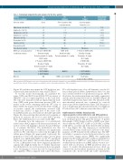
Table 1. Hematologic characteristics and outcomes for the three patients.
Patients
(age at presentation)
Infection at birth
White blood cells (x109/L) Lymphocytes (x109/L)
B lymphocytes (x109/L)
T lymphocytes (x109/L) Monocytes (x109/L) Neutrophils (x109L) Platelets (x109/L) Hemoglobin (g/dL)
Bone marrow aspirate
HSCT (age at transplantation) conditioning regimen
Donor cells
Outcome
P1 (3 days)
Sepsis
0.6
0.4
0.09
0.24
0.01
0,2
248
18
Hypoplasia
1st T-depleted HSCT (3M) Busulfan 8 mg/kg Cyclophosphamide 200 mg/kg ALS 25 mg/kg
2nd T-depleted HSCT (6M) Busulfan 16 mg/kg Cyclophosphamide 200 mg/kg ALS 25 mg/kg
1st HSCT: MMFD/f
2nd HSCT: MMFD/f
A/W
P2 (10 days)
Colored amniotic fluid, sepsis/pneumonia
0.3
0.1
0.004
0.07
NE
NE
220
13
Hypoplasia
HSCT (2 M) Busulfan 8 mg/kg Cyclophosphamide 2,000 mg/kg
MMFD/f
GvHD resolved at D120; A/W
P3 (9 days)
Sepsis/meningitis, brain abscesses
0.5
0.5
0
0
0
0
429
10
Hypoplasia
1st T-depleted HSCT (2 M) Busulfan 3.6 mg/kg Fludarabine 160 mg/m2 ALS 5 mg/kg
2nd HSCT (3M) Fludarabine 120 mg/m2 ALS 5 mg/kg
1st HSCT: MMFD/f
2nd HSCT: MMFD/f
Graft failure;
death (5.5 M post-HSCT)
Age-matched control value
7-18 3.4-7.6 0.3-2 2.5-5.5 0.1-1.1 1.5-8.5 175-500 12.5-16.6
BM: bone marrow; HSCT: hematopoietic stem cell transplantation; M: months; ALS: antilymphocyte serum; MMFD/f: mismatched family donor/father; GvHD: graft-versus-host dis- ease; A/W: alive and well (full donor immune reconstitution, and no other symptoms/diseases); NE: not evaluated.
(Figure 1D) and thus may impair the GTP hydrolysis rate, as previously demonstrated for other small GTPases.22
To test this model biochemically, we quantified the active GTP-bound RAC2 form (RAC2 GTP) in extracts of HEK293T cells not expressing RAC2 at the basal level. The cells were transduced with an empty lentiviral back- bone (WPI) with green fluorescent protein (GFP) as a tracker or the vector containing either the WT form of RAC2 cDNA, the mutated form described here (G12R) or (as positive control) the constitutively activated GTP- bound form (G12V).23 A high level of the active GTP- bound RAC2 state was observed for both G12V and G12R (Figure 1E), demonstrating that substitution by arginine at position 12 increases the level of active RAC2 protein; hence, the G12R mutation is associated with GOF.
To determine how the expression of a constitutively active form of RAC2 impacts cell division and survival, we used in vivo live cell imaging to measure the growth kinet- ics of primary fibroblasts. The proliferation rate (the change in the percentage of confluence) was significantly slower for P3’s cells than for control cells throughout the culture period (Figure 2A, upper panel). Conversely, the proportion of dead cells (measured using Cytotox green reagent) was significantly higher for the P3 experiment (Figure 2A, lower panel). These results demonstrated that, in vitro, G12R-mutated cells show impaired proliferative capacity and a high mortality rate. To further characterize the cellular defects, we used non-invasive holotomograph- ic live cell imaging to observe the cells’ biological features and organelles. When compared with control fibroblasts,
P3’s cells displayed very slow cell dynamics over the 12- hour culture period (Online Supplementary Movie V1); this was correlated with the low proliferative response. In P3’s fibroblasts, the nuclear membrane was highly segmented, and the mitochondrial network was disrupted (Figure 2B and Online Supplementary Figure S2A). The alteration of the mitochondrial network was confirmed by confocal microscopy (Online Supplementary Figure S2B); the propor- tion of fragmented mitochondrial networks was higher in P3’s fibroblasts (35%) than in control fibroblasts (10%).
G12R mutation inhibits hematopoietic stem/progenitor cell proliferation and differentiation
To understand the impact of constitutive RAC2 activa- tion on hematopoiesis and given the absence of patient BM samples, CD34+ human cord blood HSPC were trans- duced with the WPI, WT, G12R or G12V RAC2 cDNAs (with GFP as a tracker) and then cultured with cytokines for 7 days (Figure 3A). The expression of constitutively activated RAC2 forms (G12R and G12V) led to the disap- pearance of GFP+ transduced cells within 4 days. The GFP+ cell ability to generate reactive hydroxyl radical (a com- mon reactive oxygen species [ROS]) was significantly impaired in the G12R and G12V conditions (quantified as the proportion of “ROS low” cells) (Figure 3B). It is note- worthy that the addition of an ROS inducer was not asso- ciated with an elevation in ROS production (data not shown). To analyze this phenomenon in more detail, we evaluated the mitochondrial membrane potential in GFP transduced cells. In the G12R and G12V experiments,
haematologica | 2021; 106(2)
407
