
Page 29 - 2021_02-Haematologica-web
P. 29
Inherited platelet function disorders
nant transmission; other cases with a monoallelic form have mild bleeding.47,50
Biological phenotype
ADP, even at high doses, fails to induce full and irre- versible platelet aggregation. With other agonists such as thrombin receptor agonist peptide (TRAP), the snake venom protein convulxin or ristocetin, platelet aggrega- tion may remain partially reversible. A key feature is defective Giα-mediated inhibition of platelet adenylyl cyclase by ADP with loss of vasodilator-stimulated phos- phoprotein (VASP) de-phosphorylation, readily evaluated in a diagnostic test.48 Variants can result in qualitative or quantitative defects of P2Y12, the latter can also be attrib- uted to defective receptor recycling.
Genotype
The patient that we first described in 1995 has a two nucleotide deletion in the coding region of P2RY12 mov- ing the reading frame before the introduction of a prema- ture stop (p.I240fs*29).10 Subsequently described muta- tions with autosomal recessive transmission include those of the initially reported Italian family, heterozygous for two missense mutations, p.R256Q and p.R265W (Figure
3).48,49 Here, two asymptomatic family members carried the heterozygous p.R265W variant. This is important because recently a single allele p.R265P variant, affecting the same amino acid but with a different substitution, was associated with a severe phenotype and autosomal domi- nant transmission in an unrelated family (Figure 3).47 Significantly, platelet mRNA for this allele was three-fold overexpressed, limiting wild-type homodimer formation (key to ADP-receptor signaling) and introducing a domi- nant-negative effect.47 Other mutations with a single affected P2RY12 allele and mild bleeding in families with autosomal dominant transmission are shown in Figure 3.48 Missense mutations allowing normal P2Y12 synthesis and affecting domains close to or part of the ADP-binding pockets cause qualitative defects (Figure 3).48,50 Special mention should be made of a novel cytoplasmic domain p.P341A variant affecting the PDZ-binding domain and causing abnormal endosomal recycling of platelet internal pools leading to a surface deficit of P2Y12.51 Interestingly, p.R122C and p.K174E P2RY12 variants have been associ- ated with an intronic polymorphism within FR2 (encoding PAR-1) in a patient with chronic bleeding and in a patient with type 1 von Willebrand disease showing that hemor- rhage can be part of a complex trait.52,53
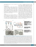
ABC
D
Figure 3. G-protein-coupled ADP receptors and signaling pathways highlighting P2RY12 variants and their different subgroups. (A) A schematic representation of
-coupled P2Y1 receptor initiates aggregation whereas the G αq
-coupled P2Y12 receptor enhances i
ADP-induced signaling pathways. Binding of extracellular ADP to the G
and sustains the platelet response. Specific signaling pathways are involved with PLCβ and PI3K enabling both RAP1 activation and RASA3 inactivation; both are required for the formation of a stable hemostatic thrombus. The release of ADP from dense granule storage pools after protein kinase C (PKC) activation contributes to the stabilization of thrombi. (B) The schematic representation shows the structure of P2Y12; variants reported as causal for a bleeding syndrome are highlighted (red spots). (C) Summarizes the genotype/phenotype relationship when the variants are regrouped according to bleeding severity and their mode of transmission. The first group consists of patients with autosomal recessive transmission and platelets showing defects of both the binding of ADP analogs and/or receptor cycling. The p.H187Q variant located in the fifth transmembrane domain has normal receptor expression but suppressed function.50 Interestingly p.R256 has a side chain that inserts into a hydrophobic pocket and in docking models it potentially makes contact with the phosphate groups of ADP;52 p.R265W impairs receptor activation and probably alters the conformational state of the receptor. Significantly, a severe phenotype-monoallelic form has a different amino acid substitution on the same residue, p.R265P associated with an increased expression of mutated mRNA and an impaired wild-type homodimer formation, possibly accounting for the bleeding severity. The third subgroup has a mild phenotype and monoallelic expression of mutations that affect receptor function: note that p.P341A located in the PDZ intra- cellular domain is associated with both abnormal ligand binding and a defect of re-sensitization of the receptor after ADP agonist-induced desensitization. (D) Electron microscopy of platelet aggregates stimulated with 10 mM ADP examined at the peak of aggregation. For the control the platelets are in close contact with some of the cells having lost their granule content. For our patient with the p.I240fs*29 mutation, platelets remain loosely bound and their granule contents are still present, as shown by the immunogold (black dots) localization of fibrinogen.
haematologica | 2021; 106(2)
343
