
Page 27 - 2021_02-Haematologica-web
P. 27
Inherited platelet function disorders
Leukocyte adhesion deficiency III syndrome
Biological phenotype and genetics
As in GT, platelets of patients with LAD-III syndrome fail to aggregate in response to all physiological agonists and PMA.30 This translates into markedly reduced throm- bus formation when whole blood is perfused over colla- gen, von Willebrand factor or fibrinogen-containing microspots.31 αIIbβ3 integrin is present but fails to bind fibrinogen or the PAC-1 monoclonal antibody when platelets are activated (Figure 2). Most disease-causing variants in FERMT3 abrogate kindlin-3 expression.29 As first shown in mice, kindlin-3 is an essential cytoplasmic cofactor for the activation of β1, β2 and β3 integrins.32 Kindlin-3 binds in a phosphorylation-dependent manner to a subterminal NITY motif of the β3 cytoplasmic tail; it coordinates with talin, which binds independently to trig- ger “inside-out” integrin activation.27,32 Initially there was controversy about the role of a CaLDAG-GEFI mutation in this disorder but this was dispelled when a splice site mutation in RASGRP2 observed in seven subjects with LAD-III syndrome was shown to be in linkage disequilib- rium with FERMT3; the two genes are located very closely on chromosome 11q.33,34 Most mutations are nonsense or involve frameshifts.
Around 2009, several groups reported that genetic vari- ants of FERMT3 encoding kindlin-3, which is expressed in all hematopoietic cells, caused leukocyte adhesion defi- ciency III (LAD-III) syndrome.29 Kindlin-3 together with its paralogs kindlin-1 and kindlin-2 are key regulators of cel- lular functions and cell-matrix interactions but kindlin-3 predominates in platelets.
Definition
LAD-III syndrome, with an autosomal recessive inheri- tance, combines a rare immunodeficiency and bleeding syndrome of extreme severity characterized by leukocyto- sis, platelet dysfunction and recurrent infections. The genetic defects result in loss of activation of β1, β2 and β3 integrins.
Clinical phenotype
Patients are detected when children have GT-like bleed- ing with severe non-purulent infections and often osteopet- rosis. The clinical phenotype is based on the description of a limited number of largely homozygous patients mostly from ethnic Turkish and Arab populations.29
A
B
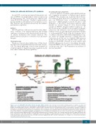
Figure 2. Loss of CalDAG-GEFI and kindlin-3 function abrogates αIIbβ3 activation. (A) A schematic representation showing examples of agonistic receptors and αIIbβ3 in the resting and activated forms. Intracytoplasmic signaling pathways induced by the binding of appropriate ligands lead to parallel roles of CalDAG-GEFI and kindlin-3 in promoting αIIbβ3 activation. CalDAG-GEFI is critical for RAP1 activation both in the circulation and at sites of vascular injury. It responds to changes in cytoplasmic Ca2+ providing sensitivity and speed to the activation response; its absence leads to a Glanzmann thrombasthenia-like phenotype. Kindlin-3 binds directly to β-integrin cytoplasmic tails in platelets but also in white blood cells, explaining the susceptibility to infections and immune disorders in its absence. (B) Key ele- ments of the syndromes resulting from RASGRP2 and FERMT3 defects are summarized. Bleeding in both cases can be very severe but defects in FERMT3 are syn- dromic and life-threatening. TRAP: thrombin receptor agonist peptid; PAR: protease activated receptor; Cvx: convulxin; GPVI: glycoprotein VI; TXA2: thromboxane A2; PLCβ/γ: phospholipase Cβ/γ; DAG: diacylglyerol: CalDAG-GEFI: calcium and diacylglycerol-regulated guanine nucleotide exchange factor I; RAP1: RAS-related protein 1; RD: related disease; AR: autosomal recessive; LTA: light transmission aggregometry; ADP: adenosine triphosphate; Col: collagen; Cvx: convulxin; AA: arachidonic acid; PMA: phorbol 12-myristate 13-acetate.
haematologica | 2021; 106(2)
341
