
Page 264 - 2021_02-Haematologica-web
P. 264
Letters to the Editor
AB
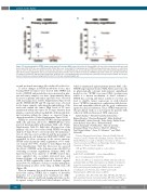
Figure 4. 5-fluorotroxacitabine (5FTRX) targets acute myeloid leukemia (AML) stem cells in vivo. (A) Primary AML cells were injected intra-femorally into irradi- ated female NOD/SCID mice preconditioned with CD122. Mice were treated with 100 mg/kg per day of 5FTRX by intraperitoneal (i.p.) injection or control from days 12-14 for 5 days (n=10 per group). Six weeks after injection, cell engraftment was assessed by flow cytometry. Data represent mean±standard deviation (SD). (B) Secondary engraftment was assessed by injecting viable leukemia cells from the bone marrow of 5FTRX treated and control mice and injected into the secondary recipient mice, which remained untreated. Six weeks later, cell engraftment was measured by flow cytometry. Data represent mean±SD. For all exper- iments, P-value was calculated using Student’s t-test; P<0.05 was considered significant.
sis and profound anti-tumor effect induced by this dose. To assess changes in 5FTRX metabolites in vivo, mice bearing MV4-11 tumors were treated with 5FTRX and levels of 5FTRX and metabolites were measured in plas- ma and tumor samples over time. Approximately linear increases in plasma and tumor exposures were observed with increasing doses (Online Supplementary Figure S13A and B). 5FTRX MP, DP and TP exposures were observed in the tumor samples, indicating phosphorylation of the nucleoside within the tumor. High levels of TP were maintained within the tumor for a longer duration than plasma, consistent with a long half-life of this metabolite, and retention within the tumor, as expected from a
charged metabolite (Online Supplementary Figure S13C). Finally, we assessed whether 5FTRX can target primary AML cells and stem cells in vivo. NOD/SCID mice were injected intra-femorally with primary AML cells (Online Supplementary Table S1). Eight days after injection of the primary cells, mice were treated with 5FTRX 100 mg/kg daily for 5 days. Four to six weeks after the initial injec- tion of cells, mice were sacrificed, and the percentage of human myeloid cells defined as CD45+CD19–CD33+ was quantified by flow cytometry in the mouse marrow. 5FTRX reduced primary AML engraftment >95% with- out toxicity (P<0.0001, Student's t-test) (Figure 4A and Online Supplementary Table S4). Moreover, we assessed the effects of 5FTRX on the leukemic stem cells (LSC) by evaluating secondary engraftment. Primary AML cells harvested from bone marrow of 5FTRX treated mice were engrafted into secondary untreated mice and human leukemic cell engraftment was assessed after 5 weeks. 5FTRX significantly reduced leukemic engraft- ment (P<0.001, Student's t-test) in secondary transplants, demonstrating an effect of the drug on AML stem cells
(Figure 4B).
Thus, in summary, this study demonstrated that the L-
nucleoside analog, 5FTRX, has a number of favorable properties for a potential new treatment for AML.1 5FTRX has a potent and broad-ranging anti-tumor activ- ity against a range of AML and other leukemic cell lines,
which is maintained against primary patient AML cells.2 5FTRX targets patient-derived AML blasts and stem cells in physiologically relevant intra-femoral engraftment models in vivo.3 5FTRX overcomes CDA overexpression which is a known mechanism of Ara-C resistance.4 5FTRX has a strong anti-tumor activity in vivo that can lead to durable tumor regressions at well-tolerated doses.5 5FTRX is synergistic in combination with doxoru- bicin and azacytidine which could be good combination agents for clinical evaluation. Thus, 5FTRX warrants fur- ther preclinical study as a novel agent for patients with refractory AML and increased CDA expression.
Aniket Bankar,1* Thirushi Piyumika Siriwardena,1*
Biljana Rizoska,2 Christina Rydergård,2 Helen Kylefjord,2 Vilma Rraklli,2 Anders Eneroth,2 Pedro Pinho,2 Stefan Norin,2 Johan Bylund,2 Sara Moses,2 Richard Bethell,2
Simon Kavanagh,1 Neil Maclean,1 Marcela Gronda,1 Xiaoming Wang,1 Rose Hurren,1 Mark D. Minden,1,3,4
Paul Targett-Adams,2 Aaron D. Schimmer1,4 and Mark Albertella2
1
Princess Margaret Cancer Center, University Health Network,
Ontario, Canada; 2Medivir AB, Huddinge, Sweden; 3Department of Medical Biophysics, Faculty of Medicine, University of Toronto, Ontario, Canada and 4Division of Hematology, Faculty of Medicine, University of Toronto, Ontario, Canada
*AB and TPS contributed equally as co-first authors.
Correspondence:
AARON D. SCHIMMER - aaron.schimmer@uhn.ca
doi:10.3324/haematol.2019.226795
Disclosures: BR was an employee of Medivir when the work was performed; MA and PTA were employees of Medivir at the time the research was performed and have an equity share in the company; ADS received research funding from Medivir and has received consulting fees from Novartis, Jazz, Takeda and Otsuka Pharmaceuticals; ADS holds shares in AbbVie.
Contributions: AB, TPS, NM, MG, RH, XW, BR, SR, PTA and MA, performed research and analyzed data; BR, MA and PTA ana- lyzed data, supervised research and provided critical reagents; ADS
578
haematologica | 2021; 106(2)
