
Page 191 - 2021_02-Haematologica-web
P. 191
ATM-induced mitophagy in MCL
AB
E
F
CD
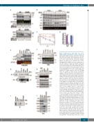
Figure 5. ATM interacts with Parkin and confers Parkin stability independent of kinase activity. (A) Immunoblot analysis (30 mg total protein) from wild- type (WT) and knocked down (Kd) HeLa cells tran- siently transfected with 3 mg of GFP-Parkin and then, 96 h later, treated with the proteasome inhibitor MG132 (10 mM) for the indicated time points. Dimethylsulfoxide (DMSO) served as a control at 0 or 12 h. Blots were cut into pieces and probed with the indicated antibodies. ATM, GFP, Parkin and GAPDH were probed using the Licor method while Pink1 (sep- arate blot from similar extract) was probed using an electrochemoluminescence (ECL) method with cells
GH treated with DMSO at 0 h as the control. GAPDH was probed as a loading control in each case. (B) ATM alters the stability of Parkin protein. Immunoblot analysis of WT and Kd HeLa cells transfected with GFP-Parkin and then, 48 h later, treated with cyclohex- imide (10 mg/mL) for the indicated times. Immunoblot analysis (30 mg total protein) from WT and Kd HeLa cells were probed with the indicated antibodies. HSP90 and Mcl-1, representing long- and short half- life proteins, were probed. Merged image showing Mcl- 1 and GAPDH (as a loading control). (C) Quantification of immunoblots from triplicate experiments as in (B). Statistical analysis was performed using a two-tailed unpaired t-test. Points represent the mean ± standard error of mean (SEM). *P≤0.05; **P≤0.01. (D) Quantitative reverse transcriptase polymerase chain reaction analysis of WT and Kd HeLa cells 72 h after transfection with GFP-Parkin, as in (B), showing the lack of effect of ATM knockdown on GFP mRNA expres- sion in either cell lines (n=3; mean ± SEM; ***P<0.001: significant difference from WT control). (E) WT and Kd HeLa cells were transiently transfected IJ with 3 μg of GFP-Parkin and then 48 h later cell lysates (500 mg) were immunoprecipitated with mouse anti- ATM antibody and probed with both GFP and Parkin antibodies. WT-GFP transfected cells were treated with CCCP to induce mitophagy (50 mM for 3 h), KU60019 (10 mM for 2 h) or neocarzinostatin (NCS) (40 nM for 2 h) before immunoprecipitation. Kd HeLa cells were left untreated and served as a negative con- trol. Arrows indicate GFP or Parkin bands superim- posed in a Licor image showing their specificity. Untreated WT-GFP transfected input cell extract (10 mg) was loaded to show the specific GFP band. IgG mouse served as an isotype matched mouse IgG con- trol. (F) Input controls (5%) of the immunoprecipitation (IP) analysis from (E). Immunoblot analysis showing total and ATMSer1981phosphorylation. Blots were cut into
haematologica | 2021; 106(2)
505
