
Page 190 - 2021_02-Haematologica-web
P. 190
A. Sarkar et al.
M
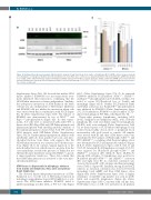
Figure 4. (Continued from the previous page). (M) Immunoblot analysis (30 μg total protein) from mantle cell lymphoma (MCL) shATM cell lines (between passage 2-5) showing loss of endogenous Parkin (electrochemoluminescence blot). Blots were cut into pieces and probed with the indicated antibodies. GAPDH was probed as a loading control. (N) Densitometry analysis showing basal Parkin expression from three separate replicates of MCL control and shATM clones. Data represent mean ± standard error of mean (n=3 from separate passages of cell lines until passage 5), *P<0.05, **P<0.01, ****P<0.0001: significant differences from respec- tive controls, as indicated.
N
Supplementary Figure S6A). We showed that neither ATM kinase inhibitor (KU60019) nor neocarzinostatin treat- ment influenced their interaction, confirming that the ATM-Parkin interaction is kinase-independent. Similarly, the endogenous interaction of ATM-Parkin in the Mino cell line was also independent of ATM kinase inhibition and KU60019 did not inhibit the interaction when cells were treated with this in combination with FCCP (Figure 5G, H; Online Supplementary Figure S6B). The efficacy of KU60019 was demonstrated by loss of ATMSer1981 and Kap1Ser824 phosphorylation (Figure 6D). In other experi- ments, A-T cells were co-transfected with either WT or kinase-dead (KD)-Flag-ATM and GFP-Parkin plasmids and immunoprecipitated with anti-Flag antibody. Analysis of the immunoprecipitates showed that both WT and Kd- ATM interacts with GFP-Parkin (Online Supplementary Figure S6C,D). Further immunoprecipitation analysis with anti-Parkin antibody from purified cytosolic and mito- chondrial fractions from Mino cells suggests that the ATM-Parkin interaction was restricted in both these frac- tions (Figure 5I, J). In contrast, trypsin digestion of the mitochondrial fraction, which removes proteins from the outer membrane, revealed the presence of Parkin but not ATM in the inner membrane along with TIM23,36 a specif- ic inner membrane translocase. These data confirm that the ATM-Parkin interaction is restricted to the outer mito- chondrial membrane.
ATM kinase is dispensable in mitophagy: evidence from mantle cell lymphoma, HeLa cells and primary B-cell lymphomas
The observed kinase-independent interaction of ATM- Parkin led us to test the role of ATM kinase function in mitophagy using pharmacological ATM inhibitors or acti- vators. The ATM kinase inhibitor, KU60019, failed to inhibit mitophagy in either MCL or WT A-T cells (Figure
6A-C; Online Supplementary Figure S7A, B). As expected KU60019 inhibited FCCP-induced ATMSer1981, H2AXSer139 and Kap1Ser824 phosphorylation in both MCL cell lines, but failed to rescue FCCP-induced loss of Tom20 and mitophagy (Figure 6D-F). Neither FCCP-induced Pink1 and Parkin activation, nor Parkin-UBSer65 phosphorylation was inhibited by KU60019 (Online Supplementary Figure S7C, D). Furthermore, KU60019 could not inhibit CCCP- induced mitophagy in WT HeLa cells (Figure 6G, H).
Thirty-eight primary lymphomas, including MCL (n=21), marginal zone lymphoma (MZL, n=5), follicular lymphoma (FL, n=6) and diffuse large B-cell lymphoma (DLBCL, n=6), were analyzed (Online Supplementary Table S2) for their response to FCCP-induced mitophagy. B cells isolated from healthy donors (n=3) or peripheral blood mononuclear cells (n=2) served as controls. All samples were treated with IR and their ATM kinase status was determined by FCS analysis of PE-phospho-ATMSer1981 and FITC-γH2AXSer139 co-staining, while their mitophagy status was determined by FCS and immunoblot analyses (Figure 6I-K, P; Online Supplementary Figures S8A-E, S9A, D and S10A-H). Among all B-cell lymphomas screened, 13 (34%) were negative for IR-induced phospho-ATMSer1981 and γH2AXSer139 activation (IR¯) (MCL=8; FL=2; DLBCL=3). Among MCL subtypes, 13 lymphomas were positive for IR-induced phospho-ATMSer1981 and γH2AXSer139 activation (IR+) and eight were IR¯; no statistical correlation was observed between IR+ or IR¯ subtypes and their mitophagy status (Table 1; Figure 6K). Similarly, among all non-MCL subjects, no statistical correlation was observed between mitophagy, basal mROS and their ATM kinase status (Figure 6M; Online Supplementary Figure S9A). A higher abundance of mtDNA copy number was seen in all IR¯ MCL and non-MCL lymphomas (Figure 6L; Online Supplementary Figure S9B). Consistent with cell line data,
was prevalent in all primary
FCCP-induced loss of DΨ m
504
haematologica | 2021; 106(2)
