
Page 186 - 2021_02-Haematologica-web
P. 186
A. Sarkar et al.
H
I
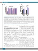
Figure 2. Defective mitophagy in mantle cell lymphoma shATM cell lines. (A) Immunoblot analysis of stable lentiviral knockdown of ATM in Jeko-1 and Mino cells showing irradiation (IR)-induced defective phospho-ATMSer1981, -Kap1Ser824, -Smc1Ser966 and -p53Ser15 expression. Separate blots (20 mg total protein) were cut into pieces and probed with the indicated antibodies. Total and phosphorylated bands were merged and are shown in color for specificity. GAPDH was probed as a loading control each time. (B) Representative flow cytometry (FCS) profile showing IR (as in Figure 1G)-induced ATMSer1981 and H2AXSer139 phosphorylation in control and shATM mantle cell lymphoma (MCL) cell lines. (C) Representative FCS profile of MitoSOX Red-stained cells showing basal levels of mitochondrial reactive oxygen species (mROS) in control and shATM MCL cell lines. (D) Relative geometric mean of mROS levels in basal and untreated (as in Figure 1F) MCL shATM clones (n=3; mean ± standard error of mean [SEM]; *P<0.05) showing significant difference from respective control shRNA. (E) Representative FCS profile (as in Figure 1A) showing mitochondrial
) in control or shATM clones treated with dimethylsulfoxide (DMSO) or FCCP in MCL cell lines. (F) Immunoblot analy- sis of whole cell extracts (30 μg total protein) from Jeko-1 and Mino shATM clones treated with FCCP or DMSO. Separate blots were cut into pieces and probed with the indicated antibodies. GAPDH was probed as a loading control each time. (G) Tom20 densitometry analysis from triplicate experiments (Figure 2F) with data pre- sented as mean ± SEM; *P<0.05, **P<0.01: significant differences from respective DMSO controls. (H) Relative geometric mean of mitochondrial mass in DMSO- or FCCP-treated (as in Figure 1B) MCL control and shATM clones (n=4; mean ± SEM). ***P<0.001, ****P<0.0001: significant differences from respective DMSO controls. (I) Quantitative polymerase chain reaction analysis of mitochondrial DNA copy number in untreated shRNA control and shATM MCL clones showing mean ±
mass and mitochondrial membrane potential (DΨ
SEM (=3). *P<0.05, **P<0.001, ****P<0.0001: significant differences from respective control shRNA. mtDNA: mitochondrial DNA.
m
autophagy resulted from the loss of functional ATM in Granta-519 cells.
Stable knockdown of ATM elicits high mitochondrial DNA copy number and defective mitophagy in mantle cell lymphoma cell lines
To further delineate the role of ATM in mitophagy, we stably knocked down ATM in Jeko-1 and Mino cells via lentiviral shATM transduction. Immunoblot analysis con- firmed loss of ATM protein in the shATM clones com- pared to control shRNA and were also defective in IR- induced ATMSer1981, Kap1Ser824, Smc1Ser966 and p53Ser15 phos- phorylation as well as IR-induced ATMSer1981 and γH2AXSer139 phosphorylation, as shown by FCS analysis (Figure 2A, B).
This decline in ATP-linked respiration in the shATM
clones resulted in depletion in the ATP pool, basal oxygen
consumption rate (OCR) or in their respective stress-
induced spare respiratory capacities (SRC) (Figure 3B-D).
The maximal ATP output of mitochondria can be deter-
These shATM clones were also found to produce rela- tively higher mROS (Figure 2C, D), defective in FCCP- induced mitophagy and retained higher mitochondrial VDAC1, Tom20, COXIV and mtDNA copy number as compared to control shRNA. Neither, autophagy (LC3 lip-
and triggers maximal OCR and substrate oxidation by complex IV. Moreover, SRC allows us to determine the ability of the cells to respond to increased energy demands following FCCP treatment. Thus, the observed decreases in both basal OCR and SRC in shATM clones were con- sistent with significant reductions in the intracellular ATP pool in ATM-depleted cells. Consistent with these find- ings, we found higher OCR and SRC in WT cells than in A-T cells (Online Supplementary Figure S4B,C).
ATM interacts with and confers Parkin stability in a kinase-independent manner
Having observed that ATM ablation impedes mitophagy prompted us to determine the role of ATM kinase in Parkin-mediated mitophagy in HeLa cells. ATM in WT HeLa cells was stably knocked down (Kd-ATM HeLa) by lentiviral shATM infection (Figure 4A). The dis- tributions of global, nuclear, cytoplasmic, and mitochon- drial ATM foci were quantified by confocal analysis using a cellular masking method (Online Supplementary Figure S5A). As expected, significantly more abundant ATM foci were observed in all three compartments in WT cells than in Kd-ATM cells (Figure 4B-D). CCCP treatment resulted in greater mitochondrial ATM-Tom20 co-localization than in control cells treated with dimethylsulfoxide. While both global and cytoplasmic ATM foci remained
idation) nor FCCP-induced loss of DΨ m
was affected by ATM ablation (Figure 2E-I and Online Supplementary Figure S3A-C). Therefore, loss of ATM provokes accumulation of mROS, preservation of mitochondrial mass, and inhibi-
tion of mitophagy in human cancer cells.
Loss of ATM is associated with less mitochondrial ATP generation
Endogenous levels of ATP and UTP were significantly higher in Jeko-1 and Mino cells (Online Supplementary Figure S4A) than in Granta-519, indicating higher respira- tory capacity. Further, ATM ablation in MCL cell lines resulted in significantly less ATP production (Figure 3A).
mined by addition of the mitochondrial uncoupler, FCCP,
which collapses the proton gradient and generates DΨ m
500
haematologica | 2021; 106(2)
