
Page 184 - 2021_02-Haematologica-web
P. 184
A. Sarkar et al.
FH
G
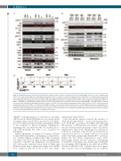
Figure 1. (Continued from the previous page) (F) Immunoblot analysis of MCL cells lines (30 mg total protein) treated with DMSO or FCCP, as in (A) or irradiation (IR) (5x10⁶ cells; 5 Gray) showing disparities in mitophagy. Separate blots were cut into pieces and probed with the indicated antibodies. Total and phosphorylated bands were merged and shown in color for specificity. Phospho-ATM, Smc-1, Kap-1 and p53 proteins represent DNA damage sensors. Decreases in Tom20 and COXIV levels reflect mitophagy and LC3 lipidation represents global autophagy. GAPDH was probed as a loading control each time. (G) FCS analysis showing IR (5 Gray)-induced ATMSer1981 and H2AXSer139 phosphorylation in MCL cell lines. One hour after IR treatment, cells were washed and stained with PE-ATMSer1981 and FITC-H2AXSer139, washed and acquired by a FACSCalibur and analyzed by FlowJo software, as in (1A). (H) Cell fractionation immunoblot analysis of MCL cell lines (30x106 cells per treatment) showing basal and FCCP (75 mM for 3 h)-induced mitophagy from whole cells (20 mg) and mitochondrial fractions (10 mg). GAPDH was probed to distinguish whole cell and mitochondrial proteins. Nuclear contamination in the mitochondrial fraction was detected by Lamin. Separate sets of blots were probed with the indicated antibodies. Parkin, phospho-Parkin and Pink1 were probed using an electrochemoluminescence method. Total and phosphorylated bands were merged and shown in color for specificity. GAPDH was probed as a loading control each time.
γH2AXSer139 phosphorylations in both Jeko-1 and Mino cells but not in Granta-519 (Figure 1G). In contrast, global autophagy induced by FCCP, as detected by LC3 lipida- tion, was similar in all three cell lines indicating that ATM is not required for global autophagy. Furthermore, mitophagy was not affected by the loss of p53 in Jeko-1 cells demonstrating that p53 is not required for mitophagy.
Similar experiments on human A-T isogenic cell lines revealed ATM dependency of mitophagy, as reported ear- lier.23 Further analyses confirmed that while autophagy was induced by CCCP in either cell line, mitophagy was restricted to only WT cells. Furthermore, we showed that A-T cells possess relatively lower levels of Pink1 and Parkin proteins with a significantly higher basal mROS level compared with the levels in WT cells (Online
Supplementary Figure S1B-F).
Cell fractionation analysis revealed the presence of
ATM and phospho-ATMSer1981 in the mitochondrial frac- tion of both Jeko-1 and Mino cells (Figure 1H). Although the majority of ATM protein was nuclear, some ATM was also detected in the cytoplasm of these cells (Online Supplementary Figure S2A). Mitophagy was detected in both Jeko-1 and Mino cells, but not in Granta-519, as evidenced by the loss of Tom20, and was associated with both Parkin and phospho-Parkin-UBSer65 activation (Figure 1H and Online Supplementary Figure S2B). Interestingly, despite selective induction of mitophagy in both Jeko-1 and Mino cells, neither p62 recruitment nor autophagy (LC3 lipidation) was affected in ATM- deficient Granta-519 cells. Together, these data suggest that a specific defect in mitophagy, but not in global
498
haematologica | 2021; 106(2)
