
Page 129 - 2021_02-Haematologica-web
P. 129
Biomarkers in Gaucher disease
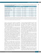
Table 2. One-stage paired comparisons of AUC-ROC curves for chitotriosidase activity and CCL18 concentration for discriminating type I Gaucher disease patients with prespecified outcomes.
Outcome
Primary composite outcome†
Secondary composite outcome‡ Hemoglobin concentration < 11 g/dL Hemoglobin concentration < 8 g/dL Platelet count < 100x109/L
Platelet count < 50x109/L Liver volume > 1.25 MN Liver volume > 2.5 MN Spleen volume > 5 MN Spleen volume > 15 MN
Symptomatic bone events#
AUC (95% CI)* n/N Chitotriosidase activity
Difference in AUC (95% CI)* P*
280/492 0.82
101/492 0.82 102/1,036 0.75 7/1,036 0.81 313/1,071 0.74 113/1,071 0.71 240/687 0.79 9/687 0.90 265/505 0.82 71/505 0.86
12/218 0.67
(0.77.- 0.87)
(0.72 - 0.89) (0.65 - 0.82) (0.58 - 0.95) (0.68 - 0.79) (0.60 - 0.80) (0.74 - 0.84) (0.81 - 0.95) (0.77 - 0.87) (0.77 - 0.92)
(0.40 - 0.83)
CCL18
0.84 (0.79 - 0.88)
0.83 (0.74 - 0.89)
0.78 (0.69 - 0.84)
0.64 (0.25 - 0.77)
0.76 (0.71 - 0.81)
0.74 (0.64 - 0.82)
0.80 (0.76 - 0.84)
0.86 (0.77 - 0.93)
0.85 (0.80 - 0.89)
0.86 (0.79 - 0.92)
0.71 (0.40 - 0.88)
0.02
0.01
0.02
-0.17
0.02
0.02
0.01
-0.04
0.03
0.00
0.04
(-0.02 to 0.05) 0.32
(-0.04 to 0.05) 0.66 (-0.04 to 0.11) 0.53
(-0.33 to -0.08)
(-0.02 to 0.06) 0.41 (-0.03 to 0.10) 0.50 (-0.02 to 0.05) 0.55 (-0.11 to 0.05) 0.34
(0.00 to 0.06) 0.09 (-0.05 to 0.06) 0.92 (-0.06 to 0.14) 0.45
0.008
AUC-ROC: area under the curve receiver operating characteristics; CI:Confidence Interval;MN:multiple of normal.*Summary estimates for area under ROC curves and P-values for paired comparisons were derived from non-parametric ROC analysis with bootstrap resampling that accounted for observation clustering within patients and primary stud- ies.†The primary outcome was a composite of hemoglobin concentration < 11 g/dL (< 10 g/dL for patients 12 to 59 months of age),platelet count < 100x109/L,spleen volume > 5 MN,and liver volume > 1.25 MN.Patients with splenectomy were excluded from this analysis.‡The secondary outcome was a composite of hemoglobin concentration < 8 g/dL (< 7 g/dL for patients 12 to 59 months of age), platelet count < 50x109/L, spleen > 15 MN, and liver volume > 2.5 MN. Patients with splenectomy were excluded from this analysis. #Osteonecrosis or fracture with imaging confirmation within the previous 12 months.
accuracy of serum CCL18 concentration in identifying patients with the primary outcome, as shown by the unchanged AUC-ROC curve. This negative finding sug- gests that combining chitotriosidase activity and serum CCL18 concentration would have a limited incremental value, probably because they convey redundant informa- tion on Gaucher cell burden.14,49-51
The recent glucosylsphingosine biomarker9 may provide complementary information about the pathophysiological process in GD.14,33 Indeed, plasma glucosylsphingosine relates to sphingolipid turnover while chitotriosidase activ- ity and CCL18 concentration are indicative of overall Gaucher cell mass. Glucosylsphingosine appeared to be highly sensitive and specific for the primary diagnosis and severity assessment of GD in previous studies.33,52 It would be interesting to compare the accuracy of glucosylsphingo- sine and CCL18 concentration using the same approach as in our meta-analysis although insufficient data exist to per- form this analysis to date. Meanwhile a prospective com- parative study of the three biomarkers is warranted to eval- uate their contribution in monitoring GD activity.
In contrast to CCL18, glucosylsphingosine can be meas- ured in dried blood spot and has emerged as a second-tier biomarker for newborn screening for GD.53 Measuring bio- markers in dried blood spot samples might facilitate GD activity monitoring for clinics where the technology is not available.
A limitation of our primary composite outcome is that it did not capture bone manifestations, despite their major impact on the quality of life.2,24 Previous studies reported that chitotriosidase activity level correlates with bone com- plications,13 history of osteonecrosis,17 and number of anatomical sites of osteonecrosis,17 although these associa- tions were inconsistent.12,15 Similarly, CCL18 concentration has been inconsistently associated with history of osteonecrosis and number of anatomical sites of osteonecrosis.12,17 Unfortunately, information on sympto- matic bone events was available in only one study inclu- ded in our meta-analysis,17 and we could not reach a definitive conclusion for this outcome.
There are several potential explanations for the see-ming- ly inconsistent findings between our analysis of bone events and the study by Deegan et al.17 First, we used a stricter definition of bone manifestations, which included symptomatic major events (i.e., skeletal fracture, osteonecrosis, or avascular necrosis) that could be dated and excluded progressive alterations (i.e., osteoporosis, Erlenmeyer flask femur deformity). Second, we took into account the data temporality by including bone events that occurred within the previous 12 months of biomarker analysis while Deegan et al.17 analyzed the previous history of bone events that occurred during the lifetime. Third, the unit of analysis was different between our analysis (218 observations nested within 97 patients) and the study by Deegan et al.17 (100 patients).
Two other clinical trials included in our meta-analysis recorded bone mineral density,30,34 but they did not report clinically relevant endpoints for bone disease (i.e., skeletal fracture, osteonecrosis, or avascular necrosis). Thus, the imprecise definition of diagnostic terms, the lack of stan- dardized criteria for bone assessment, and the slow improvement in bone outcomes after specific treatment probably hamper the identification of a relationship between biomarkers and this important clinical outcome.54- 57 Another approach in assessing skeletal disease is to meas- ure residual biomarker levels after remission of visceral dis- ease (reversal of hepatomegaly in splenectomized patients or reversal of hepatosplenomegaly in intact spleen patients).
Our findings have clinical implications for GD monitor- ing. Due to the comparable accuracy of the two biomark- ers, the impact of common genetic polymorphism on chi- totriosidase activity and the analytical robustness of CCL18 assays, it should be logical to recommend measuring CCL18 concentration rather than chitotriosidase activity as a biomarker of Gaucher cell burden and visceral and hema- tologicaldamage.
Measuring chitotriosidase activity in addition to CCL18 concentration does not appear useful in the routine practice or in clinical research. Indeed, the two biomarkers are strongly correlated and convey redundant information on
haematologica | 2021; 106(2)
443
