
Page 105 - 2021_02-Haematologica-web
P. 105
MicroRNA-21 maintains HSC homeostasis
capable of controlling the homeostasis of HSC. Recently, growing evidence has suggested that miRNA play promi- nent roles in hematopoietic cells, including HSC.17,27 In this work, we showed for the first time that miR-21 is required to maintain HSC homeostasis and function by sustaining the NF-κB pathway.
As a multifaceted, non-coding RNA, miR-21 is present in multiple tissues and implicated in various physiological and pathological processes.39 Here, we observed that miR- 21 is relatively enriched in HSC in adult mouse BM, implying that this microRNA may be involved in HSC biology. As a consequence, we found that specific knock- out of miR-21 in the hematopoietic system causes an abnormal expansion of the HSC pool in the BM and spleen. Besides, mice with conditional deletion of miR-21 display a distinctly myeloid-skewed differentiation, along with a decrease of B cells. The reason might be that miR-
AB
21 deficiency affects the differentiation of HSPC, a con- cept with is consistent with the findings of a previous study.18 In addition, a previous study showed that miR 21 can inhibit the apoptosis of diffuse large B cell lymphoma cells through upregulating BCL-2.40 Thus, miR-21 may also play a direct role in regulating B-cell survival. Taken together, our results reveal that miR-21 is essential to maintain normal hematopoiesis in mice.
Recently, miR-21 has been increasingly regarded as an oncogene due to its function in promoting tumor cell pro- liferation and inhibiting apoptosis.10 In the present study, we found that miR-21 deficiency leads to a marked impairment in HSC quiescence, accompanied by increased proliferation, which is very different from the functions of miR-21 in other types of cells. Additionally, deletion of miR-21 in HSC had no effect on cell apoptosis at a steady state. These results suggest that miR-21 regu-
CD
EFG
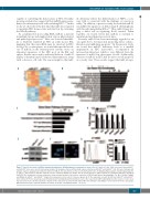
Figure 5. Specific knockout of miR-21 dramatically inhibits the NF-κB pathway in hematopoietic stem cells. (A-C) Lin-Sca1+c-Kit+ (LSK) cells sorted from miR-21∆/∆ and miR-21fl/fl mice were used for microarray analysis. (A) Heatmap of genes differentially expressed between miR-21fl/fl and miR-21∆/∆ LSK. (B) Gene Ontology (GO) analysis of genes upregulated in LSK after deletion of miR-21. The data shown are the top 15 enriched GO terms. (C) KEGG pathway analysis of downregulated genes in miR-21∆/∆ LSK relative to miR-21fl/fl LSK. The top five enriched pathways are shown. The microarray data were gathered from one experiment with three biological replicates. (D) Quantitative real-time polymerase chain reaction analysis of the relative expression of NF-κB target genes (including Egr1, Tnf, Birc3, Nr4a2, Tnfaip3 and Nfkbia) in miR-21fl/fl and miR-21∆/∆ long-term hematopoietic stem cells (n=3 mice per group). (E, F) The expression of p-p65 in LSK from miR-21fl/fl and miR-21∆/∆ bone marrow (n=5 mice per group), determined by western blotting (E) and immunofluorescence (F), respectively. DAPI staining indicates the nucleus of cells. Scale bar represents 5 mm. (G) Flow cytometric analysis of the expression of p-p65 in LSK and LT-HSC from miR-21fl/fl and miR-21∆/∆ bone marrow (n=5 mice per group). MFI: mean fluorescence intensity. All data are shown as means ± standard deviation. **P<0.01.
haematologica | 2021; 106(2)
419
