
Page 179 - 2020_11-Haematologica-web
P. 179
Letters to the Editor
A 70% cut-off for MYC protein expression in diffuse large B-cell lymphoma identifies a high-risk group of patients
We recently examined the reproducibility of MYC and BCL-2 immunohistochemical (IHC) scoring and the impact of high expression of MYC and BCL-2 (double expresser status, DE) on survival and progression in a large retrospective cohort of aggressive B-cell lymphoma patients treated with rituximab plus cyclophosphamide, doxorubicin, vincristine and prednisone (R-CHOP) or R-CHOP-like regimens.1 We found that IHC scoring for MYC and BCL-2 was highly reproducible when cut-off values of ≥70% for MYC and ≥50% for BCL-2 were used. This threshold also predicted the presence of gene rearrangements identifying MYC translocations in 88% of cases. Patients with dual MYC expression of ≥70% and BCL-2 expression of ≥50% showed a significantly inferior clinical course and, therefore, represent candi- dates for novel treatment modalities.1 We have now vali- dated these findings in an independent cohort of 461 patients enrolled in prospective clinical trials of the German High-Grade Non-Hodgkin Lymphoma Study Group (DSHNHL).2,3
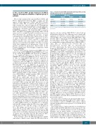
Table 1. Results from both MYC immunohistochemical (IHC) scoring and MYC fluorescence in situ hybridization.
MYC IHC
0-40%*
40%-70%*
70%-100%*
Total
MYC break
Negative
257 (97%)
96 (89%)
24 (56%)
377 (91%)
Positive
7 (3%)
12 (11%)
19 (44%)
38 (9%)
Total
264 (64%)
108 (26%)
43 (10%)
415 (100%)
In these trials, patients underwent R-CHOP-14 if >60
P<0.001. *Cut-off points were slightly different between clinical trials included in the analysis.
superior outcome and the MYC–/BCL-2+ subset had an intermediate prognosis. The differences were statistically significant for event-free survival (EFS), progression-free survival (PFS), and overall survival (OS) (EFS: DN vs. DE, P<0.001; DN vs. BCL2only P=0.004; BCL2only vs. DE P=0.032) (Figure 1A-C). These results could be confirmed in a multivariate analysis (Hazard ratios [HR] for DE vs. other: EFS: 2.1 95%CI:1.2-3.5, P=0.005; PFS: 2.5 95%CI:1.5-4.3, P=0.001; OS: 2.7 95%CI:1.5-4.8, P=0.001) adjusted for the factors of the International Prognostic Index (IPI) (age > 60 years, lactate dehydroge- nase [LDH]>N, Eastern Cooperative Oncology Group [ECOG]>1, stage III/IV, extralymphatic involvement >1). In multivariate analyses adjusted for the International Prognostic Index (IPI) factors (age > 60 years, LDH>N, ECOG>1, stage III/IV and more than one site of extra- lymphatic involvement) both MYC (70/71-100% vs. other) and BCL2 (50/51-100% vs. other) expression were significant risk factors in EFS (MYC: HR1.9, 95%CI: 1.2-3.1, P=0.007 and BCL2: HR1.8, 95%CI: 1.2-2.7, P=0.006), PFS (MYC: HR2.1, 95%CI: 1.3-3.5, P=0.004 and BCL2: HR2.4, 95%CI: 1.5-3.8, P<0.001) and OS (MYC: HR2.3, 95%CI: 1.3-4.0, P=0.004 and BCL2: HR2.0, 95%CI: 1.2-3.3 and P=0.009). When cases were stratified according to MYC protein expression only, patients with MYC ≥70%, again, experienced inferior outcome in EFS (P=0.005), PFS (P=0.004), and OS (P=0.002) in comparison with patients with low MYC expression (≤40%) (Figure 1D-F), while no difference in prognosis was seen between patients whose tumors had MYC expression ≤40% and >40-70%. Within the DE group, the occurrence of a genetic double hit for MYC and BCL-2 (n=8 of 32, 25%) failed to confer a significant prognostic difference in EFS (P=0.628), PFS (P=0.375), and OS (P=0.059) between patients with DH positive and DH negative tumors (Figure 2A-C). Within the non- DE group, we observed a genetic double hit for MYC and BCL-2 in only 11 of 354 (3%) patients with no relevant survival differences between patients with DH positive and DH negative tumors (Figure 2D-F). However, due to the low number of events, these results have to be inter- preted with caution.
In essence, these results are in agreement with our pre- vious findings indicating that high (≥70%) MYC expres- sion identifies a subset of DLBCL with adverse clinical outcome independent of the presence of a double hit of MYC and BCL-2.
Increasing evidence suggests that the sole identification of the double hit (MYC and BCL-2) status may not be the optimal tool to identify patients in need of alternative therapies and in many studies, a proportion of DE patients nevertheless experience long-term survival. Two recent papers shed light on this seeming discrepancy.8,9 In the first paper, the authors defined a clinically and biolog- ically distinct subgroup of aggressive lymphomas with
years of age and R-CHOEP/R-MegaCHOEP if ≤60 years
of age. In the MegaCHOEP trial reported by Schmitz et
al.,4 no significant differences in outcome between R-
CHOEP-14 and R-MegaCHOEP had been observed, but
to date, no randomized trial has been conducted to
answer if R-CHOEP in younger patients is superior in
comparison with R-CHOP. In a subgroup analysis for
young low-risk patients from the MInT trial reported by
Pfreundschuh et al.,5 no difference in outcome was
observed between R-CHOEP-21 and R-CHOP-21. In eld-
erly patients, the Cunningham trial6 revealed that the
outcome of R-CHOP-14 is not better than that of
R-CHOP-21. In the German cohort of 428 patients with
MYC and BCL-2 IHC scoring available, 104 cases (24%)
were MYC–/BCL-2– (double negative, DN), 283 (66%) –+
were MYC /BCL-2 (BCL2only), 8 (2%) were
+– ++
MYC /BCL-2 (MYConly), and 33 were MYC /BCL-2
using the above-mentioned cut-off values, meaning that
8% of DLBCL were assigned a DE status. Results from
both MYC IHC scoring and MYC fluorescence in situ
hybridization (FISH) were available from samples of 415
patients. In this analysis, 19 of 43 (44%) samples with
high MYC expression (70/71-100%) harbored a MYC
translocation (Table 1). The lower number of cases noted
in our report with both high MYC expression and MYC
breakage in comparison with the Ambrosio paper1 are
not easily explained. Most probably, this is due to a dif-
ference in the genetic constitution of the two different
patient populations that were examined or to the analysis
strategy: in the German cohort, the analysis was made on
1
TMA while in the paper of Ambrosio et al., full sections 7
were analyzed. According to the results of molecular cell of origin (COO) analysis, we identified 50% of patients with an ABC subtype within the DE cohort using a MYC cut-off point of 70% and 68% using a cut-off point of 40%. The sample sizes, however, are too small to con- clude that the groups differ from the proportion of the ABC subtype. It has to be stressed, however, that the DE status does not identify a homogeneous biological group of tumors and, especially, that the DE status in ABC- DLBCL arises through very different mechanisms.
In the German cohort, the DE subgroup had a signifi- cant inferior clinical course, while the DN subset had a
haematologica | 2020; 105(11)
2667
