
Page 144 - 2020_09-Haematologica-web
P. 144
M. Seibold et al.
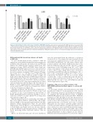
Figure 5. Combined RAL-knockdown and blockade of PI3K/Akt or MEK/MAPK signaling. L-363 cells were transfected with shRNA-expression vectors against RALA or RALB, purified next day by selection for strongly transfected cells, and then treated with PD0325901 (1 μM), MK2206 (1 μM) or BYL-719 (2.5 μM). Cell survival was measured by annexin V/PI staining after 2 days (= day 3 post-transfection). Combination of RALA knockdown with PD0325901, BYL-719 or MK-2206 treatment did not further enhance the already strong apoptosis-induction resulting from RALA depletion alone. RALB knockdown in combination with PD0325901 showed only slight additional apoptosis-induction, whereas in combination with BYL-719 or MK-2206 treatment, the rate of cell death was strongly enhanced and matched that achieved by RALA knockdown.
RNAi-mediated RAL knockdown induces cell death in MM cells
To assess whether RAL proteins contribute to MM cell survival we used an RNAi-mediated knockdown approach in HMCL with subsequent cell death assays and Western analysis. Two different target sequences against each of the respective isoforms, RALA and RALB, were cloned into pSUPER-type shRNA expression vectors. Cell survival was quantified 3 and 4 days after transfection by flow cytome- try and assessment of annexin V-FITC-negative/ PI-nega- tive events. In 4 of 7 cell lines tested, and with both shRNA-constructs, RALA depletion yielded stronger cell death effects than knockdown of RALB (Figure 2A-B and Online Supplementary Figure S2). Specifically, for cell line L- 363, viability decreased to below 40% (day 3 post-transfec- tion) and to below 30% (day 4 post-transfection) of control cells after RALA knockdown (Figure 2A). Knockdown of RALB, too, led to significantly decreased viability, albeit to a lesser extent (63-71% at day 3 post-transfection, 39-59% at day 4 post-transfection (Figure 2A).
Similarly, in MM.1S cells, knockdown of RALA led to significantly reduced cell survival to 57-64% at day 3 and to 32-42% at day 4 post-transfection. Knockdown of RALB significantly induced apoptosis leading to cell sur- vival rates of 69-87% at 3 days and 52-79% at 4 days after electroporation (Figure 2B).
RAL knockdown also led to cell death in other MM cell lines tested (INA-6, KMS-11, KMS-12-BM, and U-266), whereas AMO-1 cells remained largely unaffected by RAL depletion (Online Supplementary Figure S2 and Online Supplementary Table S3). Of note, concomitant knockdown of both RALA and RALB (tested in cell lines MM.1S, L-363, and INA-6) resulted in rapid and near complete cell death, precluding further functional analyses (data not shown).
Effects on cell metabolism and cell cycle distribution
were less pronounced than the induction of apoptosis described above. The Alamar Blue mitochondrial activity assay showed a significant decrease to 64% in L-363 cells after RALA knockdown, but only minor effects were found for MM.1S cells (Online Supplementary Figure S3A). Likewise, RALA knockdown in L-363 cells led to a signif- icant increase of the G2/M-phase from 16% to 27% after 2 days at the expense of the S-phase (decreased from 36% to 21%). After 3 days, similar effects were observed for both RALA and RALB knockdown (G2/M-phase: 20% > 24% or 25%, respectively; S-phase: 34% > 24% or 22%, respectively). For MM.1S cells, the most notable change occurred after 3 days, at which time point the share of cells in S-phase had decreased from 20 % to 14% after RALA knockdown, and to 10% after RALB knockdown (Online Supplementary Figure S3B).
Targeting of RAL does not affect activity of the MEK/MAPK pathway but RALA appears to sustain AKT activity
To investigate whether cell death induction after RAL knockdown is linked to down-regulation of the classical RAS downstream apoptosis and proliferation pathways, we analyzed the phosphorylation levels of ERK1/2 (MEK/MAPK pathway) and of Akt and GSK-3 (PI3K/Akt pathway) in L-363 and MM.1S cells after knockdown of either RALA or RALB by Western blotting. Cells were har- vested at day 2 after transfection, i.e. before the onset of significant amounts of cell death, and at day 3, at which time-point care was taken to perform sample collection such that equivalent numbers of trypan-blue negative cells were collected for control and RAL-knockdown samples. RALA or RALB depletion had no discernible effect on the phosphorylation levels of any of the above-mentioned sig- naling intermediates at day 2 (Figures 2C-D), whereas RALA knockdown specifically led to lower levels of phos-
2322
haematologica | 2020; 105(9)
