
Page 143 - 2020_09-Haematologica-web
P. 143
RAL GTPases mediate multiple myeloma cell survival
A
B
C
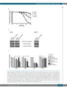
Figure 4. Pharmacological RAL inhibition in multiple myeloma cell lines. (A) AMO-1, INA-6, L-363 and MM.1S cells were subjected to increasing concentrations of the RAL inhibitor RBC8. Cell survival was measured by annexin V/PI staining after 72 hours (h) of treatment. RBC8 treatment reduced cell survival of AMO-1 and INA-6 at concentrations higher 10 μM. In contrast, L-363 and MM.1S cells showed no sensitivity towards RBC8 treatment with concentrations up to 20 μM. (B) Effect of RBC8 treatment on RAL activation status was tested in INA-6 and MM.1S cells. INA-6 cells were treated with 10 μM and 20 μM of RBC8 for 3 h, MM.1S cell were treated with 20 μM of RBC8. RAL activation assays were performed subsequently. RALA total load served as loading control in addition to a-tubulin. RAL-GTP levels were not influenced by treatment with RBC8 in MM.1S. In INA-6 20 μM of RBC8 reduced the amount of RAL-GTP compared to DMSO-treated cells, while RAL-GTP lev- els of cells treated with 10 μM of RBC remained unchanged. (C) Combined blockade of RAL and PI3K/Akt or MEK/MAPK signaling. MM cell lines (n=4) were treated for 72 h with 10 μM of RBC8, 1 μM of PD0325901, 10 μM (AMO-1, INA-6, L-363) or 2.5 μM (MM.1S) of BYL-719, 10 μM (AMO-1, L-363) or 5 μM (INA-6, MM.1S) of Akti-1,2 and the combination of RBC8 with one of the other drugs. In AMO-1 and INA-6 combination of RAL-blockade by RBC8 with blockade of MEK, PI3K or Akt1,2 by PD0325901, BYL-719 or Akti-1,2, respectively, led to a significant reduction in cell survival. MM.1S and L-363 showed no stronger decrease in cell survival after combination of RAL blockade with MEK, PI3K or Akt. Cell viability was measured with annexin V/PI staining. Bar charts show mean values and standard deviation (SD) from three independent experiments. Percentages were calculated relative to DMSO-treated control.
haematologica | 2020; 105(9)
2321
