
Page 105 - 2020_09-Haematologica-web
P. 105
SETDB1 expression suppresses MLL-fusion driven AML
SETDB1 in regulating MLL-AF9 targets, we performed an overlap analysis for genes that, upon SETDB1 overexpres- sion, are downregulated, lose promoter H3K9ac, compact chromatin, lose gene body H3K79me2 signal, and are bound by MLL-AF9.36 This defined a target list of genes that may be coregulated by SETDB1 and MLL-AF9. Included in this group is Gfi1, which has been shown to affect AML cell growth;43 Rap1gds1, a nucleotide exchange factor; Arid1b, a member of the SWI/SNF complex; and Six1, which promotes formation of leukemic stem cells40 (Figure 7C). Six1 is of particular interest given its role in promoting leukemogenesis and the striking reductions observed in H3K9ac, H3K79me2, ATAC-seq signal, and gene expression at this locus (Figure 7D). We investigated H3K9me3 levels at specific loci by performing ChIP-qPCR at the promoter of Six1 and Dock1. We detected increased H3K9me3 in MLL-AF9 cells overexpressing SETDB1
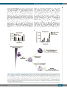
(Figure 7E). Additionally, ENCODE data from human K562 cells shows SETDB1 binding at SIX1 (Figure 7F), GFI1 (Figure S7D), RAP1GDS1, and ARID1B (data not shown), but not DOCK1 (data not shown), suggesting we identified both direct and indirect targets. To determine whether SETDB1 expression inhibits cell growth primari- ly through Hoxa9 repression, we asked whether HOXA9/MEIS1 driven colony formation is affected by SETDB1 overexpression. A modest but insignificant decrease in colony formation is observed in HOXA9/MEIS1 transformed cells following SETDB1 overexpression (Figure 8A). However, SETDB1 overex- pression is accompanied by significant upregulation of exogenous Meis1 (Figure 8B and Online Supplementary Figure S8A-B) that may account for the modest effects. Thus, Hoxa9, Meis1, and their downstream targets are likely affected by SETDB1 to influence AML cell growth.
Figure 8. SETDB1 affects Hoxa9, Meis1 and downstream targets. (A) Mouse Lin- bone marrow was retrovirally transduced with the indicated plasmid vectors and plated in methylcellulose. Colonies were counted after 7 days and re-plated, for a total of three rounds. Shown is one representative experiment of n=3, error bars are standard deviations of the technical replicates. (B) Flow cytometry showing differences in mean fluorescence intensities in HOXA9/MEIS1 transformed cells that are overexpressing SETDB1 or empty vector (EV) control. (C) Working model for the proposed role of SETDB1/H3K9 methylation in acute myeloid leukemia (AML) ini- tiation and maintenance. AML initiates from hematopoietic stem and progenitor cell (HSPC) with variable H3K9 methylation and maintain lower SETDB1 expression. After establishment of AML, inhibition of H3K9 methyltransferases leads to loss of retroviral silencing and cell death. Stabilization of SETDB1 or G9a leads to repressed Hox gene expression and relief of blocked differentiation. Statistics: generalized linear modeling followed by ANOVA where each MA9+SETDB1 replicate was paired to the MA9+EV control from the same biological replicate. Main effect is reported if there are no significant interactions (A) (See statistical analysis in the Online Supplementary Materials and Methods).
haematologica | 2020; 105(9)
2283
AB
C
