
Page 96 - Haematologica Atlas of Hematologic Cytology
P. 96
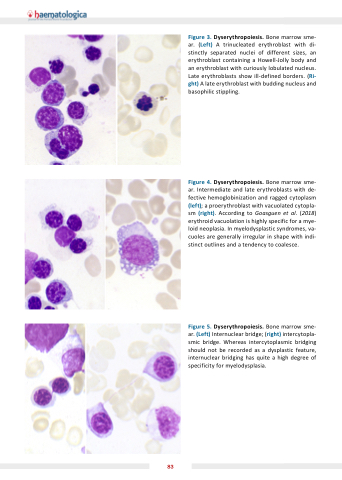
Figure Dyserythropoiesis Bone marrow sme- ar (Left) A trinucleated erythroblast with di- stinctly separated nuclei of different sizes an erythroblast containing a a a a Howell-Jolly body and an erythroblast with curiously lobulated nucleus Late erythroblasts show ill-defined borders (Ri- ght) A late erythroblast with budding nucleus and basophilic stippling Figure 4 Dyserythropoiesis Bone marrow sme- ar Intermediate and late erythroblasts with de- fective hemoglobinization and ragged cytoplasm (left) a a a a a proerythroblast with vacuolated cytopla- sm (right) According to Goasguen et al (2018) erythroid vacuolation is highly specific for a a a mye- loid neoplasia In myelodysplastic syndromes
va- cuoles are generally irregular in in shape with indi- stinct outlines and a a a tendency to coalesce Figure Dyserythropoiesis Bone marrow sme- ar ar (Left) Internuclear bridge (right) intercytopla- smic smic bridge Whereas intercytoplasmic bridging should not be recorded as as a a a a dysplastic feature internuclear bridging has quite a a a high degree of specificity for myelodysplasia 83
