
Page 67 - Haematologica Atlas of Hematologic Cytology
P. 67
CHAPTER 8 - Myeloproliferative neoplasms
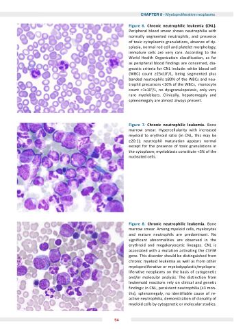
Figure hronic neutrophilic leu emia ( L) Peripheral blood smear shows neutrophilia with normally segmented neutrophils and presence of of toxic cytoplasmic granulations absence of of dy- splasia normal red cell and platelet morphology immature cells are are very rare According to the World Health Organization classification as as far as peripheral blood findings are concerned dia- gnostic criteria for CNL include: white blood cell (WBC) count ≥25x109/L being segmented plus banded neutrophils ≥80% of the WBCs and and neu- trophil precursors <10% of the WBCs monocyte count <1x10⁹/L no dysgranulopoiesis only very rare myeloblasts Clinically hepatomegaly and splenomegaly are almost always present Figure hronic neutrophilic leu emia Bone marrow smear Hypercellularity with increased myeloid to erythroid ratio (in CNL this may be ≥20:1) neutrophil maturation appears normal except for the presence of toxic granulations in the the cytoplasm myeloblasts constitute <5% of the the nucleated cells Figure hronic neutrophilic leu emia Bone marrow smear Among myeloid cells myelocytes and mature neutrophils are predominant No significant abnormalities are observed in the erythroid and megakaryocytic lineages CNL is associated with a a a a a a mutation activating the CSF3R gene This disorder should be distinguished from chronic myeloid leukemia as as well as as from other myeloproliferative or myelodysplastic/myelopro- liferative neoplasms
on the basis of cytogenetic and/or molecular analysis The distinction from leukemoid reactions rely on on clinical and genetic findings: in in in CNL persistent neutrophilia (≥3 mon- ths) splenomegaly no no identifiable cause of re- active neutrophilia demonstration of of clonality of of myeloid cells by cytogenetic or molecular studies 54
