Page 231 - Haematologica Atlas of Hematologic Cytology
P. 231
CHAPTER 27 - Acquired hemolytic anemias
Chapter 27. ACQUIRED HEMOLYTIC ANEMIAS
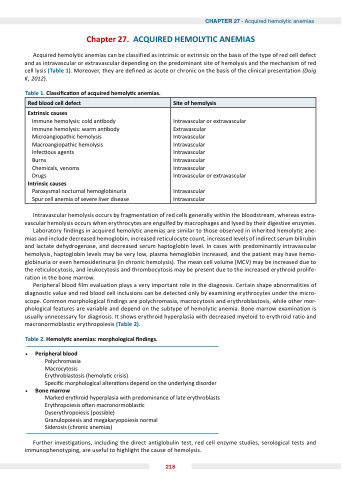
Acquired hemolytic anemias can be classified as intrinsic or extrinsic on the basis of the type of red cell defect and as intravascular or extravascular depending on the predominant site of hemolysis and the mechanism of red cell lysis (Table 1). Moreover, they are defined as acute or chronic on the basis of the clinical presentation (Doig K, 2012).
Table 1.  lassi ca on of ac uired hemoly c anemias.
Red blood cell defect
Site of hemolysis
 xtrinsic causes
Immune hemolysis: cold an body Immune hemolysis: warm an body Microangiopathic hemolysis Macroangiopathic hemolysis Infec ous agents
Burns
Chemicals, venoms Drugs
Intrinsic causes
Paroxysmal nocturnal hemoglobinuria Spur cell anemia of severe liver disease
Intravascular or extravascular Extravascular
Intravascular
Intravascular
Intravascular
Intravascular
Intravascular
Intravascular or extravascular
Intravascular Intravascular
Intravascular hemolysis occurs by fragmentation of red cells generally within the bloodstream, whereas extra- vascular hemolysis occurs when erythrocytes are engulfed by macrophages and lysed by their digestive enzymes. Laboratory findings in acquired hemolytic anemias are similar to those observed in inherited hemolytic ane- mias and include decreased hemoglobin, increased reticulocyte count, increased levels of indirect serum bilirubin and lactate dehydrogenase, and decreased serum haptoglobin level. In cases with predominantly intravascular hemolysis, haptoglobin levels may be very low, plasma hemoglobin increased, and the patient may have hemo- globinuria or even hemosiderinuria (in chronic hemolysis). The mean cell volume (MCV) may be increased due to the reticulocytosis, and leukocytosis and thrombocytosis may be present due to the increased erythroid prolife-
ration in the bone marrow.
Peripheral blood film evaluation plays a very important role in the diagnosis. Certain shape abnormalities of
diagnostic value and red blood cell inclusions can be detected only by examining erythrocytes under the micro- scope. Common morphological findings are polychromasia, macrocytosis and erythroblastosis, while other mor- phological features are variable and depend on the subtype of hemolytic anemia. Bone marrow examination is usually unnecessary for diagnosis. It shows erythroid hyperplasia with decreased myeloid to erythroid ratio and macronormoblastic erythropoiesis (Table 2).
Table 2. Hemoly c anemias  morphological  ndings.


Peripheral blood
Polychromasia
Macrocytosis
Erythroblastosis (hemoly c crisis)
Speci c morphological altera ons depend on the underlying disorder
 one marrow
Marked erythroid hyperplasia with predominance of late erythroblasts Erythropoiesis o en macronormoblas c
Dyserythropoiesis (possible)
Granulopoiesis and megakaryopoiesis normal
Siderosis (chronic anemias)
Further investigations, including the direct antiglobulin test, red cell enzyme studies, serological tests and immunophenotyping, are useful to highlight the cause of hemolysis.
218


