
Page 202 - Haematologica Atlas of Hematologic Cytology
P. 202
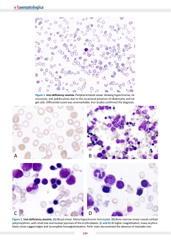
Figure 1. Iron de ciency anemia. Peripheral blood smear showing hypochromia, mi- crocytosis, and poikilocytosis due to the occasional presence of elliptocytes and tar- get cells. Di eren al count was unremarkable. Iron studies con rmed the diagnosis.
AB
CD
Figure 2. Iron de ciency anemia. (A) Blood smear. Many hypochromic microcytes. (B) Bone marrow smear reveals cellular polymorphism, with small size and nuclear pycnosis of the erythroblasts. (C and D) At higher magni ca on, many erythro- blasts show ragged edges and incomplete hemoglobiniza on. Perls  stain documented the absence of stainable iron.
189
