
Page 201 - Haematologica Atlas of Hematologic Cytology
P. 201
CHAPTER 22 - ron e cienc anemia
Chapter 22. IRON DEFICIENCY ANEMIA
Iron deficiency anemia is the most common microcytic anemia and is caused by conditions that result in iron levels insufficient for maintaining a normal hemoglobin synthesis: inadequate iron intake, increased iron requi- rement, impaired iron absorption, or chronic blood loss. Certain groups of individuals, such as menstruating wo- men, growing children, elderly subjects, soldiers, and long-distance runners are more prone to develop this type of anemia. Iron deficiency anemia develops slowly, progressing through different stages represented by storage iron depletion, functional iron depletion, and overt anemia.
Iron deficiency is suspected when hematologic findings show a hypochromic (mean corpuscular hemoglobin concentration <32 g/dL), microcytic (mean corpuscular volume <80 fL) anemia with an elevated red cell distribu- tion width (RDW). Absolute reticulocyte count is usually low. Peripheral blood smear reveals microcytosis (<6 μm in diameter) and hypochromia (increased central pallor) of red cells and the occasional presence of elliptocytes and/or target cells. White blood cell (WBC) count is usually normal and WBC differential unremarkable, while platelet count may be normal, increased or, more rarely, decreased. Diagnosis is confirmed by the results of iron studies showing decreased serum iron concentration, increased total iron binding capacity (TIBC), decreased percentage transferrin saturation, and decreased ferritin plasma concentration (Table 1). The evaluation of se- rum transferrin receptor is useful in differentiating iron deficiency anemia from anemia of chronic inflammation as the amount of circulating receptor rises when cells lack iron and decreases in chronic diseases. Erythrocyte protoporphyrin level may be higher when heme production is incomplete, but this laboratory test is not available everywhere.
Diagnosis does not usually require a bone marrow (BM) examination but this should be carried out if any doubt remains. Whereas granulocytic and megakaryocytic series are morphologically normal, erythroid precur- sors may be small in size and show nuclear pycnosis, ragged edges, and defective cytoplasmic hemoglobinization. Perls’ staining has a definitive diagnostic value as it allows tissue iron stores to be qualitatively assessed, as these are depleted or absent in iron deficiency. The presence or absence of iron stores should be evaluated by exa- mining BM macrophages in several (≥7) particles since iron is irregularly distributed in the BM. Biopsy sections are less reliable than BM aspirate and should not be used for this assessment because decalcification removes storage iron (Hughes et al., 2004).
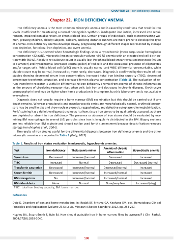
The results of iron studies useful for the differential diagnosis between iron deficiency anemia and the other microcytic anemias are reported in Table 1 (Doig, 2012).
Table 1. Results of iron status evaluation in microcytic, hypochromic anemias.
Test
Iron de ciency
Thalassemia minor
Anemia of chronic in amma on
Sideroblas c anemia
Serum iron
Decreased
Increased/normal
Decreased
Increased
TIBC
Increased
Normal
Decreased
Decreased /normal
Transferrin satura on
Decreased
Increased/normal
Decreased/normal
Increased
Serum ferri n
Decreased
Increased/normal
Increased/normal
Increased
BM storage iron
No
Increased/normal
Increased/normal
Increased
BM sideroblasts
None
Normal
None/very few
Increased (ring)
TIBC: total iron binding capacity; BM: bone marrow.
References
Doig K. Disorders of iron and heme metabolism. In: Rodak BF, Fritsma GA, Keohane EM, eds. Hematology: Clinical Principles and Applica ons (volume 2). St Louis, Missouri: Elsevier Saunders; 2012. pp. 253-267.
Hughes DA, Stuart-Smith S, Bain BJ. How should stainable iron in bone marrow lms be assessed J Clin Pathol. 2004;57(10):1038-1040.
188
