
Page 200 - Haematologica Atlas of Hematologic Cytology
P. 200
ABC
DEF
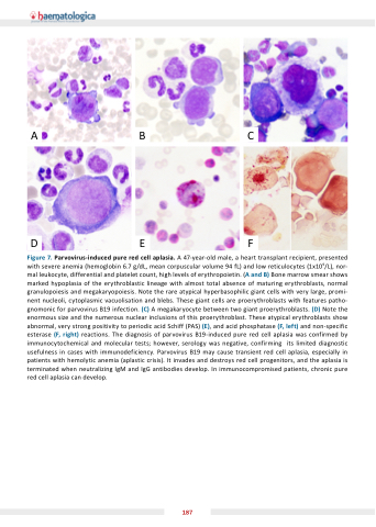
Figure 7. Parvovirus-induced pure red cell aplasia. A 47-year-old male, a heart transplant recipient, presented with severe anemia (hemoglobin 6.7 g/dL, mean corpuscular volume 94 fL) and low reticulocytes (1x10⁹/L), nor- mal leukocyte, differential and platelet count, high levels of erythropoietin. (A and B) Bone marrow smear shows marked hypoplasia of the erythroblastic lineage with almost total absence of maturing erythroblasts, normal granulopoiesis and megakaryopoiesis. Note the rare atypical hyperbasophilic giant cells with very large, promi- nent nucleoli, cytoplasmic vacuolisation and blebs. These giant cells are proerythroblasts with features patho- gnomonic for parvovirus B19 infection. (C) A megakaryocyte between two giant proerythroblasts. (D) Note the enormous size and the numerous nuclear inclusions of this proerythroblast. These atypical erythroblasts show abnormal, very strong positivity to periodic acid Schiff (PAS) (E), and acid phosphatase (F, left) and non-specific esterase (F, right) reactions. The diagnosis of parvovirus B19-induced pure red cell aplasia was confirmed by immunocytochemical and molecular tests; however, serology was negative, confirming its limited diagnostic usefulness in cases with immunodeficiency. Parvovirus B19 may cause transient red cell aplasia, especially in patients with hemolytic anemia (aplastic crisis). It invades and destroys red cell progenitors, and the aplasia is terminated when neutralizing IgM and IgG antibodies develop. In immunocompromised patients, chronic pure red cell aplasia can develop.
187
