
Page 100 - Haematologica Atlas of Hematologic Cytology
P. 100
AB
CD
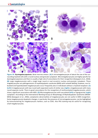
Figure 10 Dysmega aryopoiesis Bone marrow smears (A) A A micromegakaryocyte of of about the the size of of the the sur- rounding myeloid cells with a a a a a a a a round round nucleus and granular cytoplasm Micromegakaryocytes are highly specific for dysmegakaryopoiesis and there is is usually a a a a a a a a a high rate of concordance for their recognition (Goasguen et al al 2016) (B) Large megakaryocytes with a a a a a a a a a a single large round or oval eccentric nucleus and granular cytoplasm These megakaryocytes are pathognomonic of myelodysplastic syndrome (MDS) with isolated del(5q) however non-lo- bulated bulated and hypolobulated megakaryocytes occur frequently also in in individuals without a a a a a a a a a myeloid neoplasm (C) (Left) A megakaryocyte megakaryocyte with with two round well-separated nuclei of similar size (right) a a a a a a a a a megakaryocyte megakaryocyte with with many round separate nuclei There is good concordance for the recognition of multinucleated megakaryocytes which show a a a a a a a a a a significant association with with MDS (D) Micromegakaryocytes with with vacuolated (left) or hypogranular (right) cytoplasm According to to the International Working Group on on Morphology of Myelodysplastic Syndrome (IWGM- MDS) (Goasguen et al al 2016) cytoplasmic abnormalities of megakaryocytes are not useful in MDS MDS recognition Some megakaryocyte megakaryocyte dysplastic changes in particular the presence of micromegakaryocytes can be highlighted by immunostaining for for megakaryocytic markers such as CD61 Also PAS staining staining may be useful for for recognizing small megakaryocytes 87
